Math Is Fun Forum
You are not logged in.
- Topics: Active | Unanswered
- Index
- » This is Cool
- » Biopsy
Pages: 1
#1 2024-01-12 16:43:34
- Jai Ganesh
- Administrator

- Registered: 2005-06-28
- Posts: 52,688
Biopsy
Biopsy
Gist
A biopsy is a procedure to remove a piece of tissue or a sample of cells from your body so that it can be tested in a laboratory. You may undergo a biopsy if you're experiencing certain signs and symptoms or if your health care provider has identified an area of concern.
Summary
A biopsy is a medical procedure or surgery that a doctor performs to obtain a sample of cells. This can help them diagnose cancer and other health conditions that can cause abnormalities.
In some cases, your doctor may decide that he or she needs a sample of your tissue or your cells to help diagnose an illness or identify a cancer. The removal of tissue or cells for analysis is called a biopsy.
While a biopsy may sound scary, it’s important to remember that most are entirely pain-free and low-risk procedures. Depending on your situation, a piece of skin, tissue, organ, or suspected tumor will be surgically removed and sent to a lab for testing.
Why a biopsy is done
If you have been experiencing symptoms normally associated with cancer, and your doctor has located an area of concern, he or she may order a biopsy to help determine if that area is cancerous.
A biopsy is the only sure way to diagnosis most cancers. Imaging tests like CT scans and X-rays can help identify areas of concerns, but they can’t differentiate between cancerous and noncancerous cells.
Biopsies are typically associated with cancer, but just because your doctor orders a biopsy, it doesn’t mean that you have cancer. Doctors use biopsies to test whether abnormalities in your body are caused by cancer or by other conditions.
For example, if a woman has a lump in her breast, an imaging test would confirm the lump, but a biopsy is the only way to determine whether it’s breast cancer or another noncancerous condition, such as polycystic fibrosis.
Types of biopsies
There are several different kinds of biopsies. Your doctor will choose the type to use based on your condition and the area of your body that needs closer review.
Whatever the type, you’ll be given local anesthesia to numb the area where the incision is made.
Bone marrow biopsy
Inside some of your larger bones, like the hip or the femur in your leg, blood cells are produced in a spongy material called marrow.
If your doctor suspects that there are problems with your blood, you may undergo a bone marrow biopsy. This test can single out both cancerous and noncancerous conditions like leukemia, anemia, infection, or lymphoma. The test is also used to check if cancer cells from another part of the body have spread to your bones.
Bone marrow is most easily accessed using a long needle inserted into your hipbone. This may be done in a hospital or doctor’s office. The insides of your bones cannot be numbed, so some people feel a dull pain during this procedure. Others, however, only feel an initial sharp pain as the local anesthetic is injected.
Endoscopic biopsy
Endoscopic biopsies are used to reach tissue inside the body in order to gather samples from places like the bladder, colon, or lung.
During this procedure, your doctor uses a flexible thin tube called an endoscope. The endoscope has a tiny camera and a light at the end. A video monitor allows your doctor to view the images. Small surgical tools are also inserted into the endoscope. Using the video, your doctor can guide these to collect a sample.
The endoscope can be inserted through a small incision in your body, or through any opening in the body, including the mouth, nose, rectum, or urethra. Endoscopies normally take anywhere from five to 20 minutes.
This procedure can be done in a hospital or in a doctor’s office. Afterward, you might feel mildly uncomfortable, or have bloating, gas, or a sore throat. These will all pass in time, but if you are concerned, you should contact your doctor.
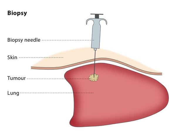
Needle biopsies
Needle biopsies are used to collect skin samples, or for any tissue that is easily accessible under the skin. The different types of needle biopsies include the following:
* Core needle biopsies use medium-sized needle to extract a column of tissue, in the same way that core samples are taken from the earth.
* Fine needle biopsies use a thin needle that is attached to a syringe, allowing fluids and cells to be drawn out.
* Image-guided biopsies are guided with imaging procedures — such as X-ray or CT scans — so your doctor can access specific areas, such as the lung, liver, or other organs.
* Vacuum-assisted biopsies use suction from a vacuum to collect cells.
Skin biopsy
If you have a rash or lesion on your skin which is suspicious for a certain condition, does not respond to therapy prescribed by your doctor, or the cause of which is unknown, your doctor may perform or order a biopsy of the involved area of skin. This can be done by using local anesthesia and removing a small piece of the area with a razor blade, a scalpel, or a small, circular blade called a “punch.” The specimen will be sent to the lab to look for evidence of conditions such as infection, cancer, and inflammation of the skin structures or blood vessels.
Surgical biopsy
Sometimes a patient may have an area of concern that cannot be safely or effectively reached using the methods described above or the results of other biopsy specimens have been negative. An example would be a tumor in the abdomen near the aorta. In this case, a surgeon may need to get a specimen using a laparoscope or by making a traditional incision.
Details
A biopsy is a medical test commonly performed by a surgeon, interventional radiologist, or an interventional cardiologist. The process involves extraction of sample cells or tissues for examination to determine the presence or extent of a disease. The tissue is then fixed, dehydrated, embedded, sectioned, stained and mounted before it is generally examined under a microscope by a pathologist; it may also be analyzed chemically. When an entire lump or suspicious area is removed, the procedure is called an excisional biopsy. An incisional biopsy or core biopsy samples a portion of the abnormal tissue without attempting to remove the entire lesion or tumor. When a sample of tissue or fluid is removed with a needle in such a way that cells are removed without preserving the histological architecture of the tissue cells, the procedure is called a needle aspiration biopsy. Biopsies are most commonly performed for insight into possible cancerous or inflammatory conditions.
History
The Arab physician Abulcasis (1013–1107) developed one of the earliest diagnostic biopsies. He used a needle to puncture a goiter and then characterized the material.
Etymology
The term biopsy reflects the Greek words bios, "life," and opsis, "a sight."
The French dermatologist Ernest Besnier introduced the word biopsie to the medical community in 1879.
Medical use:
Cancer
When cancer is suspected, a variety of biopsy techniques can be applied. An excisional biopsy is an attempt to remove an entire lesion. When the specimen is evaluated, in addition to diagnosis, the amount of uninvolved tissue around the lesion, the surgical margin of the specimen is examined to see if the disease has spread beyond the area biopsied. "Clear margins" or "negative margins" means that no disease was found at the edges of the biopsy specimen. "Positive margins" means that disease was found, and a wider excision may be needed, depending on the diagnosis.
When intact removal is not indicated for a variety of reasons, a wedge of tissue may be taken in an incisional biopsy. In some cases, a sample can be collected by devices that "bite" a sample. A variety of sizes of needle can collect tissue in the lumen (core biopsy). Smaller diameter needles collect cells and cell clusters, fine needle aspiration biopsy.
Pathologic examination of a biopsy can determine whether a lesion is benign or malignant, and can help differentiate between different types of cancer. In contrast to a biopsy that merely samples a lesion, a larger excisional specimen called a resection may come to a pathologist, typically from a surgeon attempting to eradicate a known lesion from a patient. For example, a pathologist would examine a mastectomy specimen, even if a previous nonexcisional breast biopsy had already established the diagnosis of breast cancer. Examination of the full mastectomy specimen would confirm the exact nature of the cancer (subclassification of tumor and histologic "grading") and reveal the extent of its spread (pathologic "staging").
Liquid biopsy
There are two types of liquid biopsy (which is not really a biopsy as they are blood tests that do not require a biopsy of tissue): circulating tumor cell assays or cell-free circulating tumor DNA tests. These methods provide a non-invasive alternative to repeat invasive biopsies to monitor cancer treatment, test available drugs against the circulating tumor cells, evaluate the mutations in cancer and plan individualized treatments. In addition, because cancer is a heterogeneous genetic disease, and excisional biopsies provide only a snapshot in time of some of the rapid, dynamic genetic changes occurring in tumors, liquid biopsies provide some advantages over tissue biopsy-based genomic testing. In addition, excisional biopsies are invasive, cannot be used repeatedly, and are ineffective in understanding the dynamics of tumor progression and metastasis. By detecting, quantifying and characterisation of vital circulating tumor cells or genomic alterations in CTCs and cell-free DNA in blood, liquid biopsy can provide real-time information on the stage of tumor progression, treatment effectiveness, and cancer metastasis risk. This technological development could make it possible to diagnose and manage cancer from repeated blood tests rather than from a traditional biopsy.
Circulating tumor cell tests are already available but not covered by insurance yet at maintrac and under development by many pharmaceutical companies. Those tests analyze circulating tumor cells (CTCs) Analysis of individual CTCs demonstrated a high level of heterogeneity seen at the single cell level for both protein expression and protein localization and the CTCs reflected both the primary biopsy and the changes seen in the metastatic sites.
Analysis of cell-free circulating tumor DNA (cfDNA) has an advantage over circulating tumor cells assays in that there is approximately 100 times more cell-free DNA than there is DNA in circulating tumor cells. These tests analyze fragments of tumor-cell DNA that are continuously shed by tumors into the bloodstream. Companies offering cfDNA next generation sequencing testing include Personal Genome Diagnostics and Guardant Health. These tests are moving into widespread use when a tissue biopsy has insufficient material for DNA testing or when it is not safe to do an invasive biopsy procedure, according to a recent report of results on over 15,000 advanced cancer patients sequenced with the Guardant Health test.
A 2014 study of the blood of 846 patients with 15 different types of cancer in 24 institutions was able to detect the presence of cancer DNA in the body. They found tumor DNA in the blood of more than 80 percent of patients with metastatic cancers and about 47 percent of those with localized tumors. The test does not indicate the tumor site(s) or other information about the tumor. The test did not produce false positives.
Such tests may also be useful to assess whether malignant cells remain in patients whose tumors have been surgically removed. Up to 30 percent are expected to relapse because some tumor cells remain. Initial studies identified about half the patients who later relapsed, again without false positives.
Another potential use is to track the specific DNA mutations driving a tumor. Many new cancer medications block specific molecular processes. Such tests could allow easier targeting of therapy to tumor.
Precancerous conditions
For easily detected and accessed sites, any suspicious lesions may be assessed. Originally, this was skin or superficial masses. X-ray, then later CT, MRI, and ultrasound along with endoscopy extended the range.
Inflammatory conditions
A biopsy of the temporal arteries is often performed for suspected vasculitis. In inflammatory bowel disease (Crohn's disease and ulcerative colitis), frequent biopsies are taken to assess the activity of disease and to assess changes that precede malignancy.
Biopsy specimens are often taken from part of a lesion when the cause of a disease is uncertain or its extent or exact character is in doubt. Vasculitis, for instance, is usually diagnosed on biopsy.
* Kidney disease: Biopsy and fluorescence microscopy are key in the diagnosis of alterations of renal function. The immunofluorescence plays vital role in the diagnosis of Crescentic glomerulonephritis.
* Infectious disease: Lymph node enlargement may be due to a variety of infectious or autoimmune diseases.
* Metabolic disease: Some conditions affect the whole body, but certain sites are selectively biopsied because they are easily accessed. Amyloidosis is a condition where degraded proteins accumulate in body tissues. In order to make the diagnosis, the gingival.
* Transplantation: Biopsies of transplanted organs are performed in order to determine that they are not being rejected or that the disease that necessitated transplant has not recurred.
* Fertility: A testicular biopsy is used for evaluating the fertility of men and find out the cause of a possible infertility, e.g. when sperm quality is low, but hormone levels still are within normal ranges.
Analysis of biopsied material
After the biopsy is performed, the sample of tissue that was removed from the patient is sent to the pathology laboratory. A pathologist specializes in diagnosing diseases (such as cancer) by examining tissue under a microscope. When the laboratory receives the biopsy sample, the tissue is processed and an extremely thin slice of tissue is removed from the sample and attached to a glass slide. Any remaining tissue is saved for use in later studies, if required.
The slide with the tissue attached is treated with dyes that stain the tissue, which allows the individual cells in the tissue to be seen more clearly. The slide is then given to the pathologist, who examines the tissue under a microscope, looking for any abnormal findings. The pathologist then prepares a report that lists any abnormal or important findings from the biopsy. This report is sent to the surgeon who originally performed the biopsy on the patient.
It appears to me that if one wants to make progress in mathematics, one should study the masters and not the pupils. - Niels Henrik Abel.
Nothing is better than reading and gaining more and more knowledge - Stephen William Hawking.
Offline
Pages: 1
- Index
- » This is Cool
- » Biopsy