Math Is Fun Forum
You are not logged in.
- Topics: Active | Unanswered
Pages: 1
#1 2026-03-13 18:17:05
- Jai Ganesh
- Administrator

- Registered: 2005-06-28
- Posts: 53,831
Field Vision Test
Field Vision Test
Gist
A visual field test (perimetry) maps your peripheral and central vision to detect blind spots (scotomas). It is essential for diagnosing and managing glaucoma, neurological conditions (e.g., MS, tumors, strokes), and monitoring medication side effects. Typically lasting 5–10 minutes per eye, patients click a button when they see light flashes while staring at a central point.
A visual field test can determine if you have blind spots, known as scotomas, in your vision and where they are. A blind spot's size and shape can show how eye disease or a brain disorder is affecting your vision.
Summary
A visual field test is an eye examination that can detect dysfunction in central and peripheral vision which may be caused by various medical conditions such as glaucoma, stroke, pituitary disease, brain tumours or other neurological deficits. Visual field testing can be performed clinically by keeping the subject's gaze fixed while presenting objects at various places within their visual field. Simple manual equipment can be used such as in the tangent screen test or the Amsler grid. When dedicated machinery is used it is called a perimeter.
The exam may be performed by a technician in one of several ways. The test may be performed by a technician directly, with the assistance of a machine, or completely by an automated machine. Machine-based tests aid diagnostics by allowing a detailed printout of the patient's visual field.
Details
A visual field test measures your peripheral vision, or how well you can see above, below and to the sides of something you’re looking at. It’s also called a perimetry test. Visual field testing is important for many conditions, including glaucoma.
Overview:
What is a visual field test?
A visual field test is a simple and painless test an eye care provider gives you to diagnose or monitor various eye conditions.
A visual field test measures two things:
* How far up, down, left and right your eye sees without moving (when you’re looking straight ahead).
* How sensitive your vision is in different parts of the visual field, which is the name for the entire area that you can see.
Your eyes normally see a wide area of the space in front of you. Without moving your eyes, you can see not only what’s straight ahead, but also some of what’s above, below and off to either side. Providers call all of the area you can see that isn’t right in front of you “peripheral vision.” This surrounds the area that’s right in front of you that you can see (central vision).
Vision is usually best right in the middle of the visual field, so you probably turn your eyes toward the things you want to see more clearly. The farther away from the center of your vision an object is, the less clearly you can see it. When an object moves far enough to the side, it disappears from your vision completely.
When is a visual field test performed?
When you visit an optometrist or ophthalmologist, a visual field test is part of a routine eye exam. Visual field testing can help your eye care provider find early signs of diseases like glaucoma that gradually damage vision. Some people with glaucoma don’t notice any problems with their vision, but the visual field test shows a loss of peripheral vision.
A visual field test can also help your provider find out more about the part of your nervous system that allows you to see. The visual part of your nervous system includes:
* Your retina, the part of your eye that’s like a translator that changes light energy into an electrical signal.
* Your optic nerve, the nerve that carries the signals to your brain so they can become images.
* Your brain, the place where the signals become the images you see.
Issues with any part of this system can change your visual field. There are well-known patterns in the test results that help providers recognize certain types of injury or disease.
By repeating visual field tests at regular intervals, providers also can tell whether your condition is getting better or worse.
Medical conditions that might cause a provider to order a visual field test
Your healthcare provider may want you to have a visual field test if you have (or they think you may have) certain conditions. Providers use the results to both diagnose and monitor conditions such as:
* Glaucoma.
* Stroke.
* Macular degeneration.
* Multiple sclerosis (MS).
* Graves’ disease.
* Pituitary gland disease.
* Blind spot (scotoma).
Why do some people need to have visual field tests many times?
Sometimes your eye care provider will want to repeat the visual field test right away to make sure the results are accurate. If you’re tired, for example, the test results can be unreliable.
Your provider might also recommend that you take a visual field test again in a few weeks, a few months or a year. This might be necessary to make sure that they find any new problems early. When you have certain eye conditions, your provider will do visual field tests regularly to find out how well the treatment is working.
Visual field tests are especially important in the treatment of glaucoma. These tests will tell the provider if you’re losing vision even before you notice. That’s just one of the reasons why people who have glaucoma should keep all of their appointments with their provider.
Test Details:
What happens during a visual field test?
You don’t have to prepare for a visual field test. It’s not invasive, so you aren’t likely to have any side effects.
There are several types of visual field tests, but they all have one thing in common: you look straight ahead at one point and signal when you see an object or a light somewhere off to the side.
Your provider will explain to you exactly where to look so that the test is accurate.
The two most basic types of visual field tests are very simple:
* Amsler grid: The Amsler grid is a pattern of straight lines that make perfect squares. You look at a large dot in the middle of the grid and describe any areas where the lines look blurry, wavy or broken. The Amsler grid is a quick test that only measures the middle of the visual field (your central vision) and provides your doctor with a small amount of information.
* Confrontation visual field: The term “confrontation” in this test just means that the person giving the test sits facing the person having the test, about 3 or 4 feet (around 1 meter) away. The provider holds their arms straight out to the sides. You look straight ahead, and the tester moves one hand and then the other inward toward you. You give a signal as soon as you see their hand.
The confrontation visual field test measures only the outer edge of the visual field. It’s not very exact.
Other types of visual field tests
You may hear about different types of or terms for visual field tests, including static and kinetic perimetry tests. (Perimetry test is another way of saying peripheral vision test.)
* Kinetic perimetry tests: A kinetic perimetry test is one in which the person giving the test moves an object around, and you tell them when you can see it. Providers often use the Goldmann perimetry test.
* Static perimetry tests: Automated peripheral vision tests are static perimetry tests. You look into a bowl-shaped machine and respond by pressing buttons when you see the object. Common types of static tests are the Humphrey and the Octopus.
How long does a visual field test take?
A test usually isn’t longer than about five to 10 minutes per eye.
What kind of visual field tests give more detailed information?
Computerized instruments are available to perform visual field tests and calculate results. These instruments give more reproducible and accurate results because:
* Your head is always in the same place during the test.
* The instrument has a large central “target” for you to look at, so the center of the visual field stays steady.
* The instrument uses tiny spots of light to test vision. The provider can change the brightness and color of the light to measure the sensitivity of vision at each location.
* There are clear standards for “normal” results. The instrument can compare each new test to these standards.
Results and Follow-Up:
What do the results of the visual field test mean?
A “normal” visual field test means that you can see about as well as people without vision issues.
The visual field test shows the amount of vision loss and the affected areas. The instrument prints the results as patterns of dots or numbers. The patterns tell your provider how well your eyes and visual field system work. This helps your provider diagnose an underlying health condition and what treatment you need.
A test that shows visual field loss means that vision in some areas isn’t as keen as it should be. A test could show that you have a small area of lost vision, or all vision lost in large areas.
When should I know the results of the test?
Generally, your provider should be able to give you results right away.
What are the next steps if the results are abnormal?
Abnormal results may mean different things. These results can indicate different types of issues, including glaucoma, macular degeneration or stroke. The follow-up will vary.
Your eye care provider will discuss treatment options with you.
When should I call my provider?
You should always contact your eye care provider if you have any new vision loss or eye discomfort. If you have sudden vision loss or eye pain, go to an emergency room for immediate medical help.
Additional Information
A visual field test is a diagnostic procedure that measures a person's entire field of vision, including peripheral (side) and central vision. It evaluates how well you can see in different areas of your vision and is commonly used to detect, diagnose, and monitor various eye and neurological conditions. The test plays a crucial role in identifying issues that may not be apparent during a routine eye exam, especially problems affecting peripheral vision.
Visual field testing can help uncover conditions such as glaucoma, retinal disorders, optic nerve damage, and neurological diseases like strokes or brain tumors. By mapping out the areas where vision is diminished or absent, it provides valuable insights into the health of your eyes and the visual pathways in your brain.
Importance of Test Results Interpretation
Accurate interpretation of visual field test results is critical for effective diagnosis and treatment planning. The results are presented as a detailed map showing areas where vision is normal, reduced, or absent. Key aspects of result interpretation include:
* Detection of Blind Spots: Identifying areas where vision is missing, which may indicate damage to the retina or optic nerve.
* Symmetry Analysis: Comparing the visual fields of both eyes to detect asymmetrical vision loss, which can be a sign of neurological conditions.
* Severity and Progression: Monitoring changes over time to assess the progression of diseases like glaucoma.
Patients typically receive a detailed explanation of their test results from an eye care professional, including recommendations for treatment or follow-up testing if necessary.
Uses of a Visual Field Test
Visual field tests serve a variety of purposes in both ophthalmology and neurology. Common uses include:
* Glaucoma Diagnosis and Monitoring: Identifies early signs of vision loss associated with glaucoma and tracks progression.
* Assessment of Retinal Disorders: Detects damage caused by conditions like diabetic retinopathy or retinal detachment.
* Optic Nerve Evaluation: Evaluates the health of the optic nerve, often impacted by optic neuritis or optic neuropathy.
* Neurological Conditions: Identifies vision changes due to strokes, brain tumors, or other neurological disorders.
* Pre-Surgical Planning: Assists in determining the extent of vision impairment before eye surgeries.
* Evaluation of Medication Effects: Monitors vision changes in patients taking medications that may affect eye health.
How to Prepare for a Visual Field Test
Proper preparation ensures accurate results from a visual field test. Follow these steps to get ready:
* Inform Your Eye Doctor: Share your medical history, including any eye conditions, neurological issues, or medications you are taking.
* Rest Well: Ensure you are well-rested before the test to reduce fatigue, which can affect performance.
* Wear Glasses or Contacts if Needed: Bring any corrective eyewear to the appointment, as the test may require you to wear them.
* Avoid Driving Before the Test: The procedure may involve pupil dilation, temporarily affecting your ability to drive.
* Follow Specific Instructions: Your doctor may provide additional preparation guidelines based on your individual needs.
By following these steps, you can help ensure the test provides the most accurate representation of your visual field.
What to Expect During the Procedure
A visual field test is a painless and non-invasive procedure typically performed in an eye doctor's office. Here is what what you can expect:
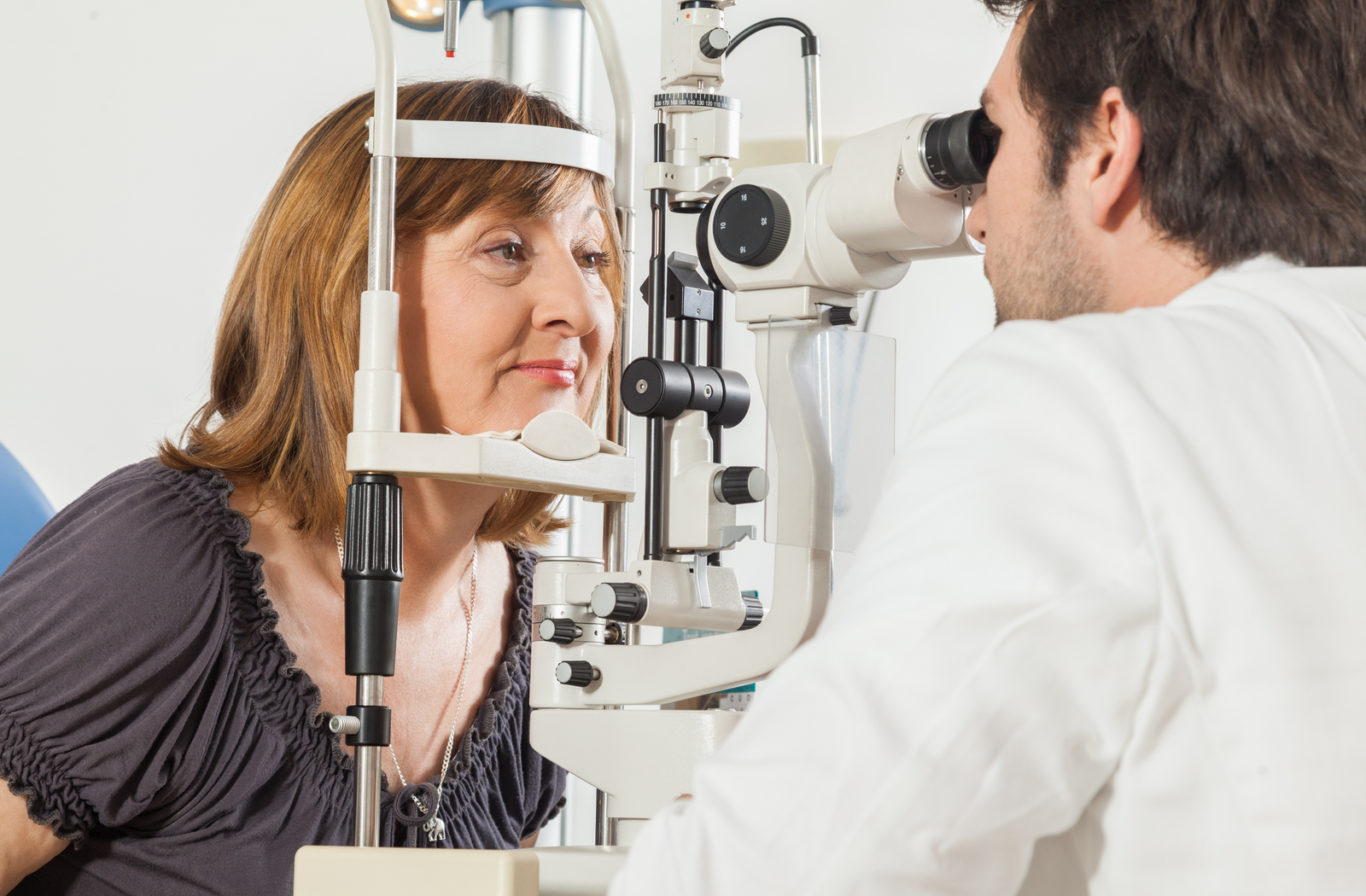
* Positioning: You will sit in front of a specialized machine and place your chin on a rest to stabilize your head.
* Focus on a Target: You will be asked to focus on a central point while small lights or objects appear in different parts of your visual field.
* Responding to Stimuli: You'll press a button or verbally indicate when you see the lights.
* Eye-by-Eye Testing: Each eye is tested separately by covering the other eye.
* Duration: The test typically takes 15-30 minutes to complete.
Patients can resume normal activities immediately after the test unless they have had their pupils dilated, in which case temporary visual sensitivity may occur.
Normal Range for Visual Field Test Results
Normal results indicate that your visual field is intact and free of significant blind spots beyond the natural blind spot (caused by the optic nerve head). Specific findings in a normal test include:
* Symmetrical vision between both eyes.
* Full peripheral vision within the expected range for your age.
* No unexplained areas of vision loss or distortion.
* Abnormal results may require further investigation to determine the underlying cause and develop an appropriate treatment plan.
Benefits of a Visual Field Test
Visual field testing offers numerous benefits for maintaining eye and neurological health. These include:
* Early Detection: Identifies vision problems before noticeable symptoms develop.
* Comprehensive Assessment: Provides a detailed map of your visual capabilities.
* Monitoring Disease Progression: Tracks changes in vision over time for conditions like glaucoma.
* Guiding Treatment Decisions: Helps tailor treatments based on the specific pattern of vision loss.
* Preventing Vision Loss: Enables timely interventions to preserve remaining vision.
Limitations and Risks of a Visual Field Test
While visual field testing is highly beneficial, it has certain limitations and risks:
* False Positives or Negatives: Patient fatigue or inattention can lead to inaccurate results.
* Limited Scope: Does not provide detailed images of the eyes' internal structures.
* Temporary Discomfort: Prolonged focus during the test may cause mild eye strain.
* Not a Standalone Diagnostic Tool: Often combined with other tests for a complete evaluation.
Understanding these limitations can help set realistic expectations for the procedure.
Frequently Asked Questions (FAQs) About Visual Field Tests:
1. Why is a visual field test important?
A visual field test is essential for detecting early signs of eye and neurological conditions, including glaucoma and optic nerve damage. It provides a detailed map of your field of vision, allowing doctors to diagnose problems that may not be noticeable during routine eye exams. Early detection through this test helps prevent further vision loss by enabling timely treatment and monitoring.
2. How often should I get a visual field test?
The frequency of visual field testing depends on your age, medical history, and risk factors. People with glaucoma or other eye conditions may need regular testing every 6-12 months. For routine eye health, adults should have a visual field test every 1-2 years as part of a comprehensive eye exam. Consult your doctor for personalized recommendations.
3. Is the visual field test painful?
No, the visual field test is completely painless and non-invasive. It involves sitting comfortably and responding to visual stimuli. Some patients may find it slightly tiring to maintain focus during the test, but there is no physical discomfort involved.
4. What do abnormal visual field test results mean?
Abnormal results indicate areas of reduced or missing vision, which could be caused by glaucoma, retinal conditions, optic nerve damage, or neurological issues like strokes. Your doctor will interpret the results and may recommend additional tests to determine the cause and guide treatment.
5. Can children undergo a visual field test?
Yes, children can undergo visual field testing if recommended by their doctor. The procedure is modified to suit their age and ability to follow instructions. It is often used to diagnose conditions like optic nerve disorders or monitor vision changes caused by neurological issues in children.
6. What is the difference between central and peripheral vision testing?
Central vision testing evaluates the ability to see details in the center of your vision, while peripheral vision testing assesses your ability to detect objects and movement in the outer areas of your field of vision. Visual field tests often include both types to provide a complete assessment.
7. Can a visual field test detect brain tumors?
Yes, a visual field test can help detect vision changes caused by brain tumors. Tumors affecting the optic pathways or visual centers in the brain can cause specific patterns of vision loss, which are identifiable through this test. Further imaging tests may be required for confirmation.
8. How accurate is a visual field test?
Visual field tests are highly accurate when performed correctly and under optimal conditions. Factors like patient attentiveness and proper calibration of the equipment influence the reliability of the results. Repeat testing may be necessary to confirm findings.
9. Are there alternatives to a visual field test?
Alternatives include fundus photography, optical coherence tomography (OCT), and perimetry tests. Each method has unique applications, and your doctor will choose the most appropriate one based on your condition and diagnostic needs.
10. What should I do if I fail a visual field test?
Failing a visual field test doesn’t always mean permanent vision loss. It indicates areas requiring further investigation. Follow your doctor’s recommendations for additional testing or treatment. Early intervention can often prevent further deterioration and improve outcomes.
Conclusion
The visual field test is an invaluable diagnostic tool for assessing and preserving eye and neurological health. By identifying early signs of vision loss and guiding treatment decisions, it plays a vital role in managing conditions like glaucoma and neurological disorders. While the procedure has certain limitations, its benefits in early detection and monitoring far outweigh them. Regular visual field testing, combined with comprehensive eye care, can help maintain optimal vision and quality of life. Consult your eye doctor to learn more about this important test and how it fits into your overall health plan.
It appears to me that if one wants to make progress in mathematics, one should study the masters and not the pupils. - Niels Henrik Abel.
Nothing is better than reading and gaining more and more knowledge - Stephen William Hawking.
Offline
Pages: 1