Math Is Fun Forum
You are not logged in.
- Topics: Active | Unanswered
Pages: 1
#1 2026-02-04 16:14:51
- Jai Ganesh
- Administrator

- Registered: 2005-06-28
- Posts: 53,783
Fetoscope/Fetoscopy
Fetoscope/Fetoscopy
Gist
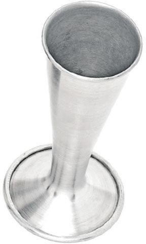
A fetoscope, also known as a fetal stethoscope or Pinard horn, is a simple, non-electronic instrument used in prenatal care to listen to a baby's heartbeat by amplifying sounds through the mother's abdomen, typically after 18-20 weeks of gestation. It's a cone-shaped device, often made of metal or plastic, that allows midwives and doctors to monitor fetal well-being without ultrasound, providing a traditional, cost-effective method for assessing the fetus.
The fetoscope allows healthcare providers, especially midwives, to monitor the heartbeat of a fetus and assess the baby's health and development. A fetal heartbeat is a vital sign that helps to detect potential issues (especially genetic conditions) early on.
Summary
A fetoscopy is a procedure that allows your healthcare team to see the inside of your uterus during pregnancy. It helps treat certain genetic conditions in a developing fetus.
Fetoscopy is a procedure during pregnancy that lets your pregnancy care provider see the fetus developing inside your uterus. Providers use it to evaluate and treat congenital disorders (diseases you’re born with). It involves inserting a thin, fiber-optic tube (endoscope or fetoscope) into your uterus through a tiny incision in your abdomen. It has a small camera on the end so your provider can see inside your uterus and amniotic sac (the sac that holds the fetus in your uterus). The fetoscope is hollow, so your provider can insert surgical tools through it, allowing them to treat certain fetal conditions or obtain samples of tissue (biopsy). In some cases, the fetoscope is inserted through your cervix instead of through your abdomen.
When is a fetoscopy done?
Fetoscopy is performed in the second or third trimester of pregnancy to treat fetal conditions or collect biopsies.
Some of the most common conditions treated with fetoscopy are:
* Twin-to-twin transfusion syndrome
Twin-to-twin transfusion syndrome is a rare, potentially life-threatening condition that occurs when identical twins aren't getting an equal share of blood while in the uterus. Your surgeon uses a fetoscope to better visualize your placenta and the blood vessels causing the condition. Then, they place a laser through the fetoscope that they use to close off the blood vessels causing uneven blood flow. This procedure is called fetoscopic laser photocoagulation.
* Amniotic band syndrome
Amniotic band syndrome occurs when the fetus gets tangled up in bands of tissue from the amniotic sac. It can restrict blood flow or cause amputation of limbs or organs. A fetoscope allows your surgeon to insert a laser device that cuts and releases the bands of tissue around the fetus.
* Congenital diaphragmatic hernia (CDH)
CDH occurs when the fetus has a hole in its diaphragm, which causes its abdominal organs to shift upward, putting pressure on the lungs. This prevents its lungs from growing properly. Surgeons use fetoscopy to insert a balloon in the fetus's airway to promote lung growth. The balloon is removed several weeks later. This procedure is called fetoscopic endoluminal tracheal occlusion (FETO).
There are other conditions fetoscopy may be used for, like treatment of placental tumors, spina bifida and other congenital diseases.
Details:
What is a Fetoscope, and Why do Midwives Use it?
A fetoscope is a medical instrument that allows healthcare providers to listen to the fetal heartbeat. Unlike a standard stethoscope, a fetoscope is designed to pick up the sounds of a baby’s heartbeat through the mother’s abdomen. It is essential to prenatal care, especially in environments prioritizing low-intervention, natural childbirth approaches, like Birthways Family Birth Center.
The Purpose of a Fetoscope
The fetoscope allows healthcare providers, especially midwives, to monitor the heartbeat of a fetus and assess the baby’s health and development. A fetal heartbeat is a vital sign that helps to detect potential issues (especially genetic conditions) early on. This monitoring is crucial throughout pregnancy, especially as the due date approaches.
Difference Between a Doppler and a Fetoscope
A fetoscope and a Doppler device detect a fetal heartbeat, but they differ in several ways:
* Technology: A Doppler uses ultrasound waves to detect the movement of the baby’s heart and translate it into sound. On the other hand, a fetoscope relies on the practitioner’s ability to manually detect and listen to the fetal heartbeat without electronic amplification.
* Sound Quality: The sound detected through a Doppler is electronically amplified to make it louder and clearer. The fetoscope provides a more natural sound but may require more skill to handle.
* Safety: The Doppler uses ultrasound waves and introduces a small amount of energy into the body. However, it is generally considered safe. A fetoscope, however, is entirely noninvasive and doesn’t involve any ultrasound or electronic waves, making it a safe option for both mother and baby.
When Can You Use it?
A fetoscope is typically used to detect a fetal heartbeat starting around 18 to 20 weeks of pregnancy. However, it requires skill and patience, as the heartbeat may not always be easy to locate, especially in early pregnancy.
Using a fetoscope is generally more effective in the later stages of pregnancy when the baby is larger and the heartbeat is stronger.
Is it Safe?
Yes, a fetoscope is entirely safe during all stages of pregnancy. It’s a non-invasive tool that does not emit radiation, ultrasound, or electronic waves. Sometimes, a fetoscope may not detect the fetal heartbeat. But this is not a concern as long as the baby is moving.
Why Do Midwives Use Them?
Midwives often prefer fetoscopes because of their safety, simplicity, and effectiveness. It aligns with the midwifery philosophy of providing natural, low-intervention care. A fetoscope creates a calm and natural environment for the mother and the baby.
To sum up, the fetoscope is a valuable tool in prenatal care. It offers a safe, effective, and natural way to monitor the baby’s health throughout pregnancy. You can learn more about fetoscopes on our YouTube channel.
Additional Information
Fetoscopy is an endoscopic procedure during pregnancy to allow surgical access to the fetus, the amniotic cavity, the umbilical cord, and the fetal side of the placenta. A small (3–4 mm) incision is made in the abdomen, and an endoscope is inserted through the abdominal wall and uterus into the amniotic cavity. Fetoscopy allows for medical interventions such as a biopsy (tissue sample) or a laser occlusion of abnormal blood vessels (such as chorioangioma) or the treatment of spina bifida.
Fetoscopy is usually performed in the second or third trimester of pregnancy. The procedure can place the fetus at increased risk of adverse outcomes, including fetal loss or preterm delivery, so the risks and benefits must be carefully weighed in order to protect the health of the mother and fetus(es). The procedure is typically performed in an operating room by an obstetrician-gynecologist.
Non-surgical fetoscopes
Fetoscopy is a surgical procedure which may involve the use of a fibreoptic device called a fetoscope. Some confusion may arise from the use of specialized forms of stethoscopes, including Pinard horns and Doppler wands, to audibly monitor fetal heart rate (FHR). These audio diagnostic tools are also called "fetoscopes" but are not related to visual fetoscopy.
It appears to me that if one wants to make progress in mathematics, one should study the masters and not the pupils. - Niels Henrik Abel.
Nothing is better than reading and gaining more and more knowledge - Stephen William Hawking.
Offline
Pages: 1