Math Is Fun Forum
You are not logged in.
- Topics: Active | Unanswered
- Index
- » Science HQ
- » Skull
Pages: 1
#1 2026-01-19 18:08:36
- Jai Ganesh
- Administrator

- Registered: 2005-06-28
- Posts: 53,783
Skull
Skull
Gist
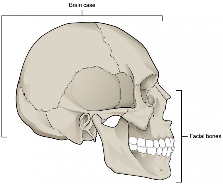
A skull is the bony structure forming the head, protecting the brain and supporting facial features, but it also symbolizes mortality, knowledge (the mind), or danger (skull and crossbones) across cultures, representing the fragility of life or a warning. It consists of cranial bones (braincase) and facial bones, connected by immobile sutures, with the movable jaw (mandible) being a key part.
The rounded brain case surrounds and protects the brain and houses the middle and inner ear structures. In the adult, the skull consists of 22 individual bones, 21 of which are immobile and united into a single unit. The 22nd bone is the mandible (lower jaw), which is the only moveable bone of the skull.
Summary
Skull is a skeletal framework of the head of vertebrates, composed of bones or cartilage, which form a unit that protects the brain and some sense organs. The upper jaw, but not the lower, is part of the skull. The human cranium, the part that contains the brain, is globular and relatively large in comparison with the face. In most other animals the facial portion of the skull, including the upper teeth and the nose, is larger than the cranium. In humans the skull is supported by the highest vertebra, called the atlas, permitting nodding motion. The atlas turns on the next-lower vertebra, the axis, to allow for side-to-side motion.
In humans the base of the cranium is the occipital bone, which has a central opening (foramen magnum) to admit the spinal cord. The parietal and temporal bones form the sides and uppermost portion of the dome of the cranium, and the frontal bone forms the forehead; the cranial floor consists of the sphenoid and ethmoid bones. The facial area includes the zygomatic, or malar, bones (cheekbones), which join with the temporal and maxillary bones to form the zygomatic arch below the eye socket; the palatine bone; and the maxillary, or upper jaw, bones. The nasal cavity is formed by the vomer and the nasal, lachrymal, and turbinate bones. In infants the sutures (joints) between the various skull elements are loose, but with age they fuse together. Many mammals, such as the dog, have a sagittal crest down the centre of the skull; this provides an extra attachment site for the temporal muscles, which close the jaws.
Details
The skull, or cranium, is typically a bony enclosure around the brain of a vertebrate. In some fish and amphibians, the skull is of cartilage. The skull is at the head end of the vertebrate.
In a human, the skull comprises two prominent parts: the neurocranium and the facial skeleton, which evolved from the first pharyngeal arch. The skull forms the frontmost portion of the axial skeleton and is a product of cephalization and vesicular enlargement of the brain, with several special senses structures such as the eyes, ears, nose, tongue and, in fish, specialized tactile organs such as barbels near the mouth.
The skull is composed of three types of bone: cranial bones, facial bones and ossicles, which is made up of a number of fused flat and irregular bones. The cranial bones are joined at firm fibrous junctions called sutures and contains many foramina, fossae, processes, and sinuses. In zoology, the openings in the skull are called fenestrae, the most prominent of which is the foramen magnum, where the brainstem goes through to join the spinal cord.
In human anatomy, the neurocranium (or braincase), is further divided into the calvaria and the endocranium, together forming a cranial cavity that houses the brain. The interior periosteum forms part of the dura mater, the facial skeleton and splanchnocranium with the mandible being its largest bone. The mandible articulates with the temporal bones of the neurocranium at the paired temporomandibular joints. The skull itself articulates with the spinal column at the atlanto-occipital joint. The human skull fully develops two years after birth.
Functions of the skull include physical protection for the brain, providing attachments for neck muscles, facial muscles and muscles of mastication, providing fixed eye sockets and outer ears (ear canals and auricles) to enable stereoscopic vision and sound localisation, forming nasal and oral cavities that allow better olfaction, taste and digestion, and contributing to phonation by acoustic resonance within the cavities and sinuses. In some animals such as ungulates and elephants, the skull also has a function in anti-predator defense and sexual selection by providing the foundation for horns, antlers and tusks.
Additional Information:
Overview
What is the skull?
Your skull is the part of your skeleton that holds and protects your brain. It also holds or supports several of your main sensory organs, like your eyes, ears, nose, tongue and more. The skull’s medical name is the cranium.
When you’re born, your skull is mostly formed but not quite complete. Some parts of it, called fontanelles, are softer and more flexible. You can see an example of this in the soft spot at the top of an infant’s head. The fontanelles allow your brain and skull to grow and develop.
There are also places where the left and right sides of a bone (like the parietal bone) or multiple bones join. Those are called sutures. These can change throughout your lifetime, even well into adulthood. Some sutures fuse solid while you’re an infant. Others may not fuse until your 60s.
Function:
What does the skull do?
Your skull has two main jobs:
* Protection. The bony structure of your skull protects your brain and critical sensory organs like your eyes and ears.
* Structure. Your skull is what gives your face and head their shape. It has many attachment points for muscles to anchor to. It’s one of the most important things that determines what your face looks like.
Anatomy:
What are the parts of the skull?
Your skull sits at the top of your spinal column, inside your neck. While it might seem like your skull is just one structure, there are two distinct parts of it. They are the:
* Cranial vault (neurocranium or calvarium). This surrounds and protects your brain.
* Facial skeleton (viscerocranium). This supports and holds the various parts of your face.
Cranial vault anatomy
This part of your skull consists mainly of the calvarium. That includes the:
* Frontal bone. This is a single, seamless bone. It gives your forehead its structure.
* Sphenoid bone. This is a single bone below and in front of your brain, but behind the bones making up your face. It also forms part of the lower rear of your eye sockets.
* Ethmoid bone. This is a single bone that fills in a heart-shaped, hollow space in the sphenoid bone at the lower front of your brain.
* Temporal bones. There are two of these bones, one on each side. Each has a small opening where your ear canal passes through the skull.
* Parietal bones. These are a pair of bones that join together at a seam called the sagittal suture. They form the upper middle and upper back of your skull.
* Occipital bone. This is a single seamless bone at the lower back of your skull.
Facial skeletal anatomy
The facial skeleton is at the front of your skull. It’s a group of bones that support and give your face its structure. The bones of this part of your skull are:
* Nasal bones. These form the bridge of your nose. They overlap slightly with the maxilla and the forward part of the ethmoid bone.
* Vomer. This bone is like the floor of the nasal cavity space right behind your nose.
* Lacrimal bones. These form the central-lower inside portion of your eye socket.
* Palatine bones. These are a small section of the bottom of your eye socket’s interior.
* Zygomatic bones. These form the outer lower edge of your eye socket.
* Maxilla. This single bone forms the central part of your cheekbones on both sides. It also is what makes up the central-lower-forward part of your eye socket. It also makes up your upper jaw. It overlaps slightly with the forward part of the ethmoid bone.
* Mandible. This single bone makes up your lower jaw. It’s the only bone of your skull that moves.
Conditions and Disorders:
What are the common conditions and disorders that affect the skull?
Your skull is prone to a wide range of conditions, and many of them are congenital. That means you have the condition when you’re born. Examples of congenital skull conditions include:
* Anencephaly
* Apert syndrome
* Carpenter syndrome
* Cleidocranial dysplasia
* Craniosynostosis
* Crouzon syndrome
* Encephalocele
* Goldenhar syndrome
* Hemifacial microsmia
* Microcephaly
* Micrognathia
* Pfeiffer syndrome
* Prognathism
* Scaphocephaly
There are also several conditions and injuries that you can develop at any time in life that affect your skull. Examples include:
* Temporomandibular joint disorders
* Jaw cysts and tumors, some of which can be jaw cancer
* Dental trauma, including a dislocated jaw or broken jaw
* Skull fractures and/or related concussions and traumatic brain injuries (TBIs)
Common signs or symptoms of skull conditions
The common signs and symptoms of skull conditions vary widely. Congenital skull conditions usually cause differences in skull appearance and/or development. That can cause distinctive facial or head appearances.
Non-congenital skull conditions can cause the following:
* Head pain, especially headaches
* Jaw popping or jaw pain
* Brain symptoms, like confusion or coma
* Bruising, often with specific patterns like raccoon eyes (a key sign of a skull fracture)
Common tests to check the skull
Diagnostic imaging tests are the main way to diagnose skull conditions. These include:
* X-rays, including dental x-rays
* CT scans
* MRI
Providers who suspect congenital conditions that affect your skull often recommend genetic testing. It can detect specific genetic variations that could help providers find the right diagnosis.
Other tests are possible, depending on your symptoms, health history and other factors. Your healthcare provider is the best person to tell you what tests they recommend and why.
What are some common treatments for skull conditions?
The treatments for skull conditions depend on which condition you have. Your health, personal history and circumstances can also be factors. Ask your healthcare provider about the treatment options for your specific case.
It appears to me that if one wants to make progress in mathematics, one should study the masters and not the pupils. - Niels Henrik Abel.
Nothing is better than reading and gaining more and more knowledge - Stephen William Hawking.
Offline
Pages: 1
- Index
- » Science HQ
- » Skull