Math Is Fun Forum
You are not logged in.
- Topics: Active | Unanswered
Pages: 1
#1 2025-10-30 22:28:26
- Jai Ganesh
- Administrator

- Registered: 2005-06-28
- Posts: 53,783
Coagulation
Coagulation
Gist
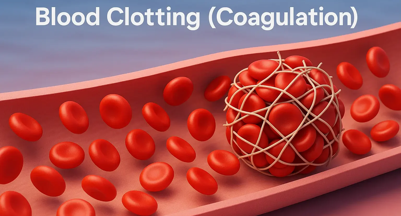
Coagulation, or blood clotting, is the process where blood changes from a liquid to a gel to form a clot, which stops bleeding when a blood vessel is injured. This process is a critical part of hemostasis, where platelets and proteins like fibrin work together to create a plug at the injury site, which is then stabilized by a fibrin mesh. Disorders of this system can lead to either excessive bleeding or dangerous, spontaneous blood clots.
Blood clotting, or coagulation, is an important process that prevents excessive bleeding when a blood vessel is injured. Platelets (a type of blood cell) and proteins in your plasma (the liquid part of blood) work together to stop the bleeding by forming a clot over the injury.
Summary
Coagulation, also known as clotting, is the process by which blood changes from a liquid to a gel, forming a blood clot. It results in hemostasis, the cessation of blood loss from a damaged vessel, followed by repair. The process of coagulation involves activation, adhesion and aggregation of platelets, as well as deposition and maturation of fibrin.
Coagulation begins almost instantly after an injury to the endothelium that lines a blood vessel. Exposure of blood to the subendothelial space initiates two processes: changes in platelets, and the exposure of subendothelial platelet tissue factor to coagulation factor VII, which ultimately leads to cross-linked fibrin formation. Platelets immediately form a plug at the site of injury; this is called primary hemostasis. Secondary hemostasis occurs simultaneously: additional coagulation factors beyond factor VII respond in a cascade to form fibrin strands, which strengthen the platelet plug.
Coagulation is highly conserved throughout biology. In all mammals, coagulation involves both cellular components (platelets) and proteinaceous components (coagulation or clotting factors). The pathway in humans has been the most extensively researched and is the best understood. Disorders of coagulation can result in problems with hemorrhage, bruising, or thrombosis.
Details:
Role in disease
Coagulation defects may cause hemorrhage or thrombosis, and occasionally both, depending on the nature of the defect.
The GP1b-IX receptor complex. This protein receptor complex is found on the surface of platelets, and in conjunction with GPV allows for platelets to adhere to the site of injury. Mutations in the genes associated with the glycoprotein Ib-IX-V complex are characteristic of Bernard–Soulier syndrome.
Platelet disorders
Platelet disorders are either congenital or acquired. Examples of congenital platelet disorders are Glanzmann's thrombasthenia, Bernard–Soulier syndrome (abnormal glycoprotein Ib-IX-V complex), gray platelet syndrome (deficient alpha granules), and delta storage pool deficiency (deficient dense granules). Most are rare. They predispose to hemorrhage. Von Willebrand disease is due to deficiency or abnormal function of von Willebrand factor, and leads to a similar bleeding pattern; its milder forms are relatively common.
Decreased platelet numbers (thrombocytopenia) is due to insufficient production (e.g., myelodysplastic syndrome or other bone marrow disorders), destruction by the immune system (immune thrombocytopenic purpura), or consumption (e.g., thrombotic thrombocytopenic purpura, hemolytic-uremic syndrome, paroxysmal nocturnal hemoglobinuria, disseminated intravascular coagulation, heparin-induced thrombocytopenia). An increase in platelet count is called thrombocytosis, which may lead to formation of thromboembolisms; however, thrombocytosis may be associated with increased risk of either thrombosis or hemorrhage in patients with myeloproliferative neoplasm.
Coagulation factor disorders
The best-known coagulation factor disorders are the hemophilias. The three main forms are hemophilia A (factor VIII deficiency), hemophilia B (factor IX deficiency or "Christmas disease") and hemophilia C (factor XI deficiency, mild bleeding tendency).
Von Willebrand disease (which behaves more like a platelet disorder except in severe cases), is the most common hereditary bleeding disorder and is characterized as being inherited autosomal recessive or dominant. In this disease, there is a defect in von Willebrand factor (vWF), which mediates the binding of glycoprotein Ib (GPIb) to collagen. This binding helps mediate the activation of platelets and formation of primary hemostasis.
In acute or chronic liver failure, there is insufficient production of coagulation factors, possibly increasing risk of bleeding during surgery.
Thrombosis is the pathological development of blood clots. These clots may break free and become mobile, forming an embolus or grow to such a size that occludes the vessel in which it developed. An embolism is said to occur when the thrombus (blood clot) becomes a mobile embolus and migrates to another part of the body, interfering with blood circulation and hence impairing organ function downstream of the occlusion. This causes ischemia and often leads to ischemic necrosis of tissue. Most cases of venous thrombosis are due to acquired states (older age, surgery, cancer, immobility). Unprovoked venous thrombosis may be related to inherited thrombophilias (e.g., factor V Leiden, antithrombin deficiency, and various other genetic deficiencies or variants), particularly in younger patients with family history of thrombosis; however, thrombotic events are more likely when acquired risk factors are superimposed on the inherited state.
Additional Information
Blood clotting, or coagulation, is an important process that prevents excessive bleeding when a blood vessel is injured. Platelets (a type of blood cell) and proteins in your plasma (the liquid part of blood) work together to stop the bleeding by forming a clot over the injury. Typically, your body will naturally dissolve the blood clot after the injury has healed. Sometimes, however, clots form on the inside of vessels without an obvious injury or do not dissolve naturally. These situations can be dangerous and require accurate diagnosis and appropriate treatment.
Clots can occur in veins or arteries, which are vessels that are part of the body's circulatory system. While both types of vessels help transport blood throughout the body, they each function differently. Veins are low-pressure vessels that carry deoxygenated blood away from the body's organs and back to the heart. An abnormal clot that forms in a vein may restrict the return of blood to the heart and can result in pain and swelling as the blood gathers behind the clot.
Blood Clots in the Arteries
Arteries, on the other hand, are muscular, high-pressure vessels that carry oxygen- and nutrient-rich blood from the heart to other parts of the body. When your doctor measures your blood pressure, the test results are an indicator of the pressure in your arteries. Clotting that occurs in arteries is usually associated with atherosclerosis (hardening of the arteries), a deposit of plaque that narrows the inside of the vessel. As the arterial passage narrows, the strong arterial muscles continue to force blood through the opening, and the high pressure can cause the plaque to rupture. Molecules released in the rupture cause the body to overreact and form an unnecessary clot in the artery, potentially leading to a heart attack or stroke. When the blood supply to the heart or brain is completely blocked by the clot, a part of these organs can be damaged as a result of being deprived of blood and its nutrients.
Coagulation, in physiology, is the process by which a blood clot is formed. The formation of a clot is often referred to as secondary hemostasis, because it forms the second stage in the process of arresting the loss of blood from a ruptured vessel. The first stage, primary hemostasis, is characterized by blood vessel constriction (vasoconstriction) and platelet aggregation at the site of vessel injury. Under abnormal circumstances, clots can also form in a vessel that has not been breached; such clots can result in the occlusion (blockage) of the vessel (see thrombosis).
Clotting is a sequential process that involves the interaction of numerous blood components called coagulation factors. There are 13 principal coagulation factors in all, and each of these has been assigned a Roman numeral, I to XIII. Coagulation can be initiated through the activation of two separate pathways, designated extrinsic and intrinsic. Both pathways result in the production of factor X. The activation of this factor marks the beginning of the so-called common pathway of coagulation, which results in the formation of a clot.
The extrinsic pathway is generally the first pathway activated in the coagulation process and is stimulated in response to a protein called tissue factor, which is expressed by cells that are normally found external to blood vessels. However, when a blood vessel breaks and these cells come into contact with blood, tissue factor activates factor VII, forming factor VIIa, which triggers a cascade of reactions that result in the rapid production of factor X. In contrast, the intrinsic pathway is activated by injury that occurs within a blood vessel. This pathway begins with the activation of factor XII (Hageman factor), which occurs when blood circulates over injured internal surfaces of vessels. Components of the intrinsic pathway also may be activated by the extrinsic pathway; for example, in addition to activating factor X, factor VIIa activates factor IX, a necessary component of the intrinsic pathway. Such cross-activation serves to amplify the coagulation process.
The production of factor X results in the cleavage of prothrombin (factor II) to thrombin (factor IIa). Thrombin, in turn, catalyzes the conversion of fibrinogen (factor I)—a soluble plasma protein—into long, sticky threads of insoluble fibrin (factor Ia). The fibrin threads form a mesh that traps platelets, blood cells, and plasma. Within minutes, the fibrin meshwork begins to contract, squeezing out its fluid contents. This process, called clot retraction, is the final step in coagulation. It yields a resilient, insoluble clot that can withstand the friction of blood flow.
It appears to me that if one wants to make progress in mathematics, one should study the masters and not the pupils. - Niels Henrik Abel.
Nothing is better than reading and gaining more and more knowledge - Stephen William Hawking.
Offline
Pages: 1