Math Is Fun Forum
You are not logged in.
- Topics: Active | Unanswered
Pages: 1
#1 2025-12-12 22:08:41
- Jai Ganesh
- Administrator

- Registered: 2005-06-28
- Posts: 53,783
Cerebellum
Cerebellum
Gist
The cerebellum, Latin for "little brain," is a crucial part of the brain located at the back, beneath the cerebrum, vital for coordinating movement, balance, posture, and motor learning, essentially fine-tuning commands from the motor cortex to ensure smooth, precise actions by detecting and correcting "motor errors". It receives sensory info and motor plans, compares intended movement with actual execution, and sends corrective signals, influencing everything from walking and talking to complex tasks, with damage leading to coordination loss and slurred speech.
The cerebellum, Latin for "little brain," is a crucial part of the hindbrain located at the back of the head, primarily responsible for coordinating voluntary movements, balance, posture, and motor learning, ensuring smooth, precise actions by fine-tuning signals from the cerebrum and brainstem; it's also increasingly recognized for roles in cognition, emotion, and social behavior.
Summary
The cerebellum (pl.: cerebella or cerebellums; Latin for 'little brain') is a major feature of the hindbrain of all vertebrates. Although usually smaller than the cerebrum, in some animals such as the mormyrid fishes it may be as large as it or even larger. In humans, the cerebellum plays an important role in motor control and cognitive functions such as attention and language as well as emotional control such as regulating fear and pleasure responses, but its movement-related functions are the most solidly established. The human cerebellum does not initiate movement, but contributes to coordination, precision, and accurate timing: it receives input from sensory systems of the spinal cord and from other parts of the brain, and integrates these inputs to fine-tune motor activity. Cerebellar damage produces disorders in fine movement, equilibrium, posture, and motor learning in humans.
Anatomically, the human cerebellum has the appearance of a separate structure attached to the bottom of the brain, tucked underneath the cerebral hemispheres. Its cortical surface is covered with finely spaced parallel grooves, in striking contrast to the broad irregular convolutions of the cerebral cortex. These parallel grooves conceal the fact that the cerebellar cortex is actually a thin, continuous layer of tissue tightly folded in the style of an accordion. Within this thin layer are several types of neurons with a highly regular arrangement, the most important being Purkinje cells and granule cells. This complex neural organization gives rise to a massive signal-processing capability, but almost all of the output from the cerebellar cortex passes through a set of small deep nuclei lying in the white matter interior of the cerebellum.
In addition to its direct role in motor control, the cerebellum is necessary for several types of motor learning, most notably learning to adjust to changes in sensorimotor relationships. Several theoretical models have been developed to explain sensorimotor calibration in terms of synaptic plasticity within the cerebellum. These models derive from those formulated by David Marr and James Albus, based on the observation that each cerebellar Purkinje cell receives two dramatically different types of input: one comprises thousands of weak inputs from the parallel fibers of the granule cells; the other is an extremely strong input from a single climbing fiber. The basic concept of the Marr–Albus theory is that the climbing fiber serves as a "teaching signal", which induces a long-lasting change in the strength of parallel fiber inputs. Observations of long-term depression in parallel fiber inputs have provided some support for theories of this type, but their validity remains controversial.
Details
Your cerebellum is part of your brain that helps coordinate and regulate a wide range of functions and processes in both your brain and body. While it’s very small compared to your brain overall, it holds more than half of the neurons (cells that make up your nervous system) in your whole body.
What is the cerebellum?
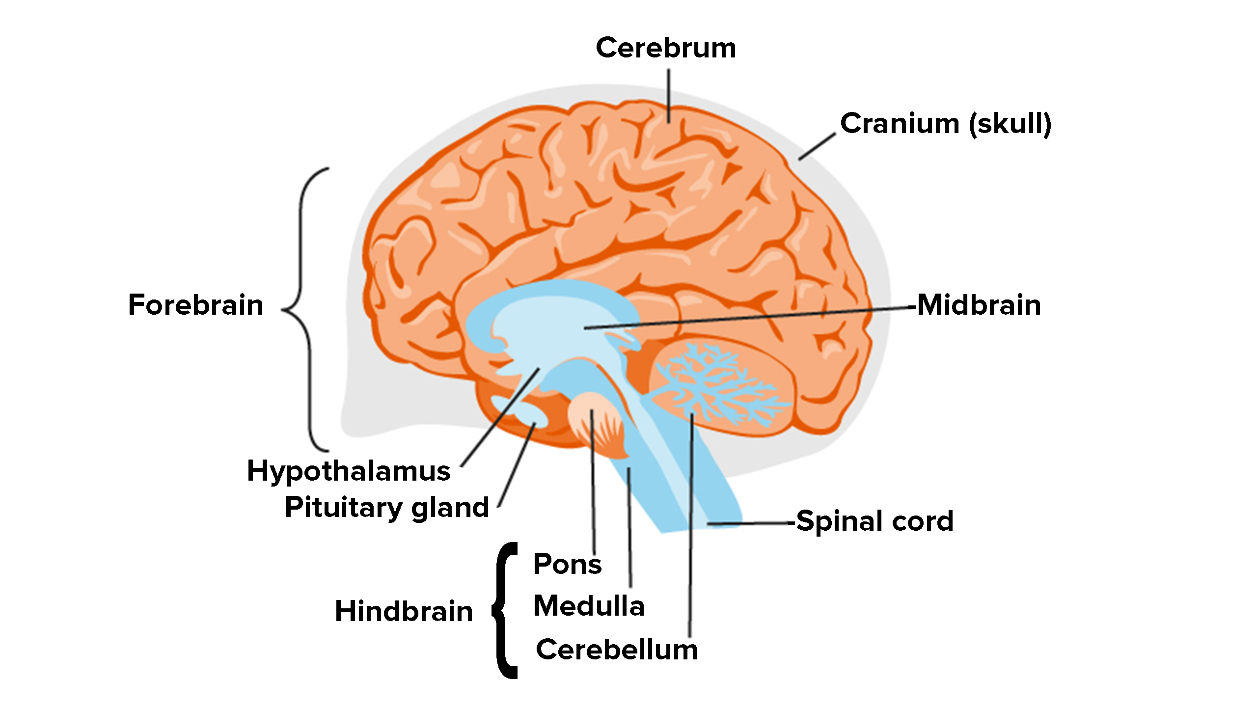
Your cerebellum is a part of your brain located at the back of your head, just above and behind where your spinal cord connects to your brain itself. The name “cerebellum” comes from Latin and means “little brain.”
For centuries, scientists believed your cerebellum’s job was to coordinate your muscle movements. Advances in technology have shown that your cerebellum does much more than that. There’s much that scientists are still trying to understand about the cerebellum, including all the ways it works with the rest of your nervous system.
What’s the difference between the cerebellum and cerebrum?
Your cerebellum is a small part of your brain located at the bottom of this organ near the back of your head. Your cerebrum is the largest part of your brain and includes parts above and forward of the cerebellum.
Function:
What does the cerebellum do?
Scientists started analyzing the cerebellum more than 200 years ago by studying people or animals with cerebellum damage. They found people with this kind of damage usually had trouble keeping their balance while standing or walking, or they’d have trouble reaching for objects because their hands would miss an object they were trying to pick up.
Over time, scientists started finding evidence that cerebellum damage could have other effects. They found that damage could make it harder or for a person to learn new words or skills. Damage to your cerebellum can interfere with judging the size of or distance from objects. It can also affect your sense of timing. As an example, people with damage to their cerebellum may have trouble repeatedly tapping their fingers, causing them to tap too soon or too late from beat-to-beat.
Advances in technology have done even more to improve experts’ understanding of the cerebellum. Now, scientists can image a person’s brain activity while that person does a certain task. What scientists have found (so far) is that different parts of your cerebellum are more active depending on what you’re doing at the time. They’ve also found that your cerebellum plays a role in emotions and how you make decisions.
Can you live without a cerebellum?
There are cases of people born with cerebellar agenesis, which is being born without a cerebellum. This condition is extremely rare. Many people with it have only minor effects. They can walk and have lives that are more or less like anyone else’s. Others have severe symptoms and will need constant medical care for their entire life.
People can also survive injuries or diseases that damage their cerebellum, but it’s common for them to have long-term or permanent issues.
What are some interesting facts about the cerebellum?
Neurons are specialized cells that make up your nervous system, including your brain, spinal cord and all of your nerves. Your cerebellum is only about 10% of your brain in terms of how much space it takes up. However, it holds about half of all the neurons in your entire body.
Your cerebellum is also incredibly compact. The brain tissue that makes up your cerebellum is a sheet folded up like an accordion. Laid flat, it would be a little over 3 feet long and 4 inches wide (1 meter by 10 centimeters).
Anatomy:
Where is the cerebellum located?
Your cerebellum is inside of your head, at about the same level as your ears. In relation to the rest of your brain, it’s at the very bottom and sits just above where your neck meets your skull.
What does it look like?
Your cerebellum forms a half-circle shape around your brain stem, which connects your brain to your spinal cord. It has a series of horizontal grooves from top to bottom.
What color is it?
Your cerebellum is a pinkish-gray color.
How big is it?
The average adult cerebellum is about 4.5 inches (11.5 centimeters) wide. In the middle, it’s between 1 inch and 1.5 inches (3 centimeters - 4 centimeters) tall. On the sides, it’s between 2 inches and 2.5 inches (5 centimeters - 6 centimeters) tall.
How much does it weigh?
The average adult cerebellum weighs between 4.8 ounces and 6 ounces (136 grams - 169 grams).
Conditions and Disorders:
What are the common conditions and disorders that affect this body system or organ?
Any condition that can affect your brain can affect your cerebellum. Some major examples include:
* Ataxia (this is both a symptom and a group of diseases).
* Congenital disorders (conditions you have at birth, such as Chiari malformation).
* Immune and inflammatory conditions (an example of this is multiple sclerosis).
* Genetic disorders (conditions you have at birth that you inherited from one or both parents, such as Wilson’s disease).
* Infections (these can happen because of bacteria, viruses, parasites and fungi).
* Vitamin deficiencies and nutrition problems (such as low vitamin B12 levels).
* Stroke.
* Cancer.
* Cerebellar agenesis (being born without a cerebellum at all).
Common signs or symptoms of conditions affecting your cerebellum?
Many symptoms can happen with conditions affecting your cerebellum. Some of the most common symptoms include:
* Dysarthria: Problems with your cerebellum can affect your ability to speak clearly.
* Ataxia: This is a loss of coordination. It can make you clumsy, causing balance problems or trouble using your hands for common tasks.
* Dizziness.
* Paralysis: This can affect various parts of your body.
* Shaking or tremors: Loss of muscle coordination can cause parts of your body, especially your hands, to shake.
* Vision problems: Your cerebellum plays a role in controlling your eyes and how your brain processes what you see. Conditions that affect your cerebellum can cause double vision (diplopia) or other problems.
Common tests to check the health of the body organ?
Many types of tests can help diagnose conditions that affect your cerebellum, including:
* Blood tests (these can look for anything from immune system problems to toxins and poisons, especially certain metals like copper).
* Genetic testing.
* Magnetic resonance imaging (MRI).
* Spinal tap (lumbar puncture).
Common treatments for the body organ?
The treatments for conditions that affect your cerebellum depend entirely on the conditions themselves. They can range from antibiotics for bacterial infections to radiation and chemotherapy for brain tumors. There’s no one-size-fits-all for treating problems that affect your cerebellum.
The cerebellum is located at the base of the brain, under the cerebrum and posterior to the spinal cord. The cerebellum is relatively small, but it is neuron-rich, containing over 50% of the brain’s neurons in a dense cellular layer, called the cerebellar cortex.
Functions of the Cerebellum
The cerebellum plays a vital role in function and mobility. Traditionally known functions of the cerebellum include:
* motor movement regulation, including gait coordination and maintenance of posture
* balance control
* control of muscle tone and voluntary muscle activity
* motor learning
There is also growing research on the cerebellum's role in emotion and cognition, specifically with visual-spatial memory, the creation of generative grammar into the structure of the brain, and the conscious ability to manipulate cause-and-effect relationships.
The cerebellum makes fine adjustments to motor actions. Four principles important to cerebellar processing have been identified.
* Feedforward processing
* Divergence and convergence
* Modularity
* Plasticity
Anatomical Position
The cerebellum is located at the back of the brain, immediately inferior to the occipital and temporal lobes, and within the posterior cranial fossa. It is separated from the occipital and temporal lobes by the tentorium cerebelli, a tough layer of dura mater. It is posterior to the pons. The fourth ventricle separates the pons from the cerebellum.
Cerebellar Structure
The cerebellum has two hemispheres. These hemispheres are connected by the vermis, a narrow midline area. The cerebellum receives input and transmits output via a limited number of cells. It is divided into thousands of independent modules, all with a similar structure.
The cerebellum is made up of grey matter and white matter:
* grey matter: located on the surface of the cerebellum. The grey matter forms the cerebellar cortex. It is tightly folded (i.e. convoluted to increase its surface area) and is divided into three layers: the molecular layer (external); the Purkinje cell layer (middle); and the granular layer (internal). There are two types of neurons in the molecular layer: the outer stellate cell and the inner basket cell.
* white matter: located below the cerebellar cortex. Four cerebellar nuclei (dentate, emboliform, globose, and fastigial nuclei) are embedded in the white matter.
The cerebellum can be subdivided in the following ways: (1) anatomical lobes, (2) zones and (3) functional divisions.
Additional Information
Cerebellum is the section of the brain that coordinates sensory input with muscular responses, located just below and behind the cerebral hemispheres and above the medulla oblongata.
The cerebellum integrates nerve impulses from the labyrinths of the ear and from positional sensors in the muscles; cerebellar signals then determine the extent and timing of contraction of individual muscle fibres to make fine adjustments in maintaining balance and posture and to produce smooth, coordinated movements of large muscle masses in voluntary motions.
Like the cerebrum, the cerebellum is divided into two lateral hemispheres, which are connected by a medial part called the vermis. Each of the hemispheres consists of a central core of white matter and a surface cortex of gray matter and is divided into three lobes. The flocculonodular lobe, the first section of cerebellum to evolve, receives sensory input from the vestibules of the ear; the anterior lobe receives sensory input from the spinal cord; and the posterior lobe, the last to evolve, receives nerve impulses from the cerebrum. All of these nerve impulses are integrated within the cerebellar cortex. Three paired bundles of nerve fibres relay information to and from the cerebellum—the superior, middle, and inferior peduncles—which connect the cerebellum with the midbrain, pons, and medulla, respectively.
Functionally, the cerebellar cortex is divided into three layers: an outer synaptic layer (also called the molecular layer), an intermediate discharge layer (the Purkinje layer), and an inner receptive layer (the granular layer). Sensory input from different types of receptors is conveyed to specific regions of the receptive layer, which is made up of numerous small nerve cells that project axons into the synaptic layer. There the axons excite the dendrites of the Purkinje cells, which in turn project axons to portions of the four intrinsic nuclei (known as the dentate, globose, emboliform, and fastigial nuclei) and upon dorsal portions of the lateral vestibular nucleus. Most Purkinje cells use the neurotransmitter GABA and therefore exert strong inhibitory influences upon the cells that receive their terminals. As a result, all sensory input into the cerebellum results in inhibitory impulses’ being exerted upon the deep cerebellar nuclei and parts of the vestibular nucleus. Cells of all deep cerebellar nuclei, on the other hand, are excitatory (secreting the neurotransmitter glutamate) and project upon parts of the thalamus, red nucleus, vestibular nuclei, and reticular formation.
Injuries or disease affecting the cerebellum usually produce neuromuscular disturbances, in particular ataxia, or disruptions of coordinated limb movements. The loss of integrated muscular control may cause tremors and difficulty in standing.
It appears to me that if one wants to make progress in mathematics, one should study the masters and not the pupils. - Niels Henrik Abel.
Nothing is better than reading and gaining more and more knowledge - Stephen William Hawking.
Offline
Pages: 1