Math Is Fun Forum
You are not logged in.
- Topics: Active | Unanswered
- Index
- » This is Cool
- » Vein
Pages: 1
#1 2025-08-12 17:53:31
- Jai Ganesh
- Administrator

- Registered: 2005-06-28
- Posts: 52,814
Vein
Vein
Gist
Vein : A blood vessel that carries blood to the heart from tissues and organs in the body.
Veins are blood vessels located throughout your body that collect oxygen-poor blood and return it to your heart. Veins are part of your circulatory system. They work together with other blood vessels and your heart to keep your blood moving. Veins hold most of the blood in your body.
Summary
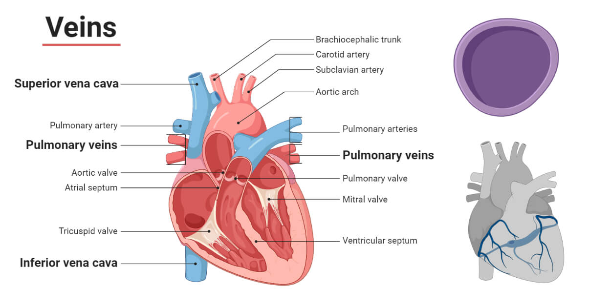
A vein, in human physiology, is any of the vessels that, with four exceptions, carry oxygen-depleted blood to the right upper chamber (atrium) of the heart. The four exceptions—the pulmonary veins—transport oxygenated blood from the lungs to the left upper chamber of the heart. The oxygen-depleted blood transported by most veins is collected from the networks of microscopic vessels called capillaries by thread-sized veins called venules.
As in the arteries, the walls of veins have three layers, or coats: an inner layer, or tunica intima; a middle layer, or tunica media; and an outer layer, or tunica adventitia. Each coat has a number of sublayers. The tunica intima differs from the inner layer of an artery: many veins, particularly in the arms and legs, have valves to prevent backflow of blood, and the elastic membrane lining the artery is absent in the vein, which consists primarily of endothelium and scant connective tissue. The tunica media, which in an artery is composed of muscle and elastic fibres, is thinner in a vein and contains less muscle and elastic tissue, and proportionately more collagen fibres (collagen, a fibrous protein, is the main supporting element in connective tissue). The outer layer (tunica adventitia) consists chiefly of connective tissue and is the thickest layer of the vein. As in arteries, there are tiny vessels called vasa vasorum that supply blood to the walls of the veins and other minute vessels that carry blood away. Veins are more numerous than arteries and have thinner walls owing to lower blood pressure. They tend to parallel the course of arteries.
Details
Veins are blood vessels in the circulatory system of humans and most other animals that carry blood towards the heart. Most veins carry deoxygenated blood from the tissues back to the heart; exceptions are those of the pulmonary and fetal circulations which carry oxygenated blood to the heart. In the systemic circulation, arteries carry oxygenated blood away from the heart, and veins return deoxygenated blood to the heart, in the deep veins.
There are three sizes of veins: large, medium, and small. Smaller veins are called venules, and the smallest the post-capillary venules are microscopic that make up the veins of the microcirculation. Veins are often closer to the skin than arteries.
Veins have less smooth muscle and connective tissue and wider internal diameters than arteries. Because of their thinner walls and wider lumens they are able to expand and hold more blood. This greater capacity gives them the term of capacitance vessels. At any time, nearly 70% of the total volume of blood in the human body is in the veins. In medium and large sized veins the flow of blood is maintained by one-way (unidirectional) venous valves to prevent backflow. In the lower limbs this is also aided by muscle pumps, also known as venous pumps that exert pressure on intramuscular veins when they contract and drive blood back to the heart.
Structure
There are three sizes of vein, large, medium, and small. Smaller veins are called venules. The smallest veins are the post-capillary venules. Veins have a similar three-layered structure to arteries. The layers known as tunicae have a concentric arrangement that forms the wall of the vessel. The outer layer, is a thick layer of connective tissue called the tunica externa or adventitia; this layer is absent in the post-capillary venules. The middle layer, consists of bands of smooth muscle and is known as the tunica media. The inner layer, is a thin lining of endothelium known as the tunica intima. The tunica media in the veins is much thinner than that in the arteries as the veins are not subject to the high systolic pressures that the arteries are. There are valves present in many veins that maintain unidirectional flow.
Unlike arteries, the precise location of veins varies among individuals.
Veins close to the surface of the skin appear blue for a variety of reasons. The factors that contribute to this alteration of color perception are related to the light-scattering properties of the skin and the processing of visual input by the visual cortex, rather than the actual colour of the venous blood which is dark red.
Venous system
The venous system is the system of veins in the systemic and pulmonary circulations that return blood to the heart. In the systemic circulation the return is of deoxygenated blood from the organs and tissues of the body, and in the pulmonary circulation the pulmonary veins return oxygenated blood from the lungs to the heart. Almost 70% of the blood in the body is in the veins, and almost 75% of this blood is in the small veins and venules. All of the systemic veins are tributaries of the largest veins, the superior and inferior vena cava, which empty the oxygen-depleted blood into the right atrium of the heart. The thin walls of the veins, and their greater internal diameters (lumens) enable them to hold a greater volume of blood, and this greater capacitance gives them the term of capacitance vessels. This characteristic also allows for the accommodation of pressure changes in the system. The whole of the venous system, bar the post-capillary venules is a large volume, low pressure system. The venous system is often asymmetric, and whilst the main veins hold a relatively constant position, unlike arteries, the precise location of veins varies among individuals.
Veins vary in size from the smallest post-capillary venules, and more muscular venules, to small veins, medium veins, and large veins. The thickness of the walls of the veins varies as to their location – in the legs the vein walls are much thicker than those in the arms. In the circulatory system, blood first enters the venous system from capillary beds where arterial blood changes to venous blood.
Large arteries such as the thoracic aorta, subclavian, femoral and popliteal arteries lie close to a single vein that drains the same region. Other arteries are often accompanied by a pair of veins held in a connective tissue sheath. The accompanying veins are known as venae comitantes, or satellite veins, and they run on either side of the artery. When an associated nerve is also enclosed, the sheath is known as a neurovascular bundle. This close proximity of the artery to the veins helps in venous return due to the pulsations in the artery. It also allows for the promotion of heat transfer from the larger arteries to the veins in a counterflow exchange that helps to preserve normal body heat.
Additional Information
Veins are blood vessels that carry oxygen-poor blood to your heart. Pulmonary veins are an exception because they carry oxygen-rich blood from your lungs to your heart. Veins in your legs fight gravity to push blood up toward your heart. Common problems with veins include chronic venous insufficiency, deep vein thrombosis and varicose veins.
What are veins?
Veins are blood vessels located throughout your body that collect oxygen-poor blood and return it to your heart. Veins are part of your circulatory system. They work together with other blood vessels and your heart to keep your blood moving. Veins hold most of the blood in your body. In fact, nearly 75% of your blood is in your veins.
What type of blood do veins carry?
The major difference between arteries and veins is the type of blood they carry. While arteries carry oxygen-rich blood, veins carry oxygen-poor blood. Your pulmonary veins are an exception to this rule. These four veins, located between your heart and lungs, carry oxygen-rich blood from your lungs back to your heart. From there, your heart pumps the oxygen-rich blood back throughout your body.
What are venules?
Your venules are very small blood vessels that connect your capillaries with your veins throughout your body. Your venules have the important function of moving blood that contains waste and lacks oxygen from your capillaries to your veins. From there, your blood can make its way back to your heart.
Your venules are wider than your capillaries but narrower than your veins. Venules vary in size, but even the widest venule is about 16 times smaller than your typical vein.
Function:
What do veins do?
Veins have two main purposes. One purpose is to collect oxygen-poor blood throughout your body and carry it back to your heart. The other purpose is to carry oxygen-rich blood from your lungs to your heart. This is the only time veins carry oxygen-rich blood.
The purpose of each vein depends upon where it’s located within your body. Veins are organized into a complex network called the venous system.
The venous system
The venous system refers to your network of veins and the way your veins connect with other blood vessels and organs throughout your body. Your venous system is organized into two main parts or circuits. These are the systemic circuit and the pulmonary circuit. Each circuit relies on blood vessels (veins, arteries and capillaries) to keep blood moving.
To help understand how these circuits work, you might think of a racetrack. At a racetrack, the race cars must complete many laps around an entire course (circuit). But the cars can’t keep going without refueling and getting quick tune-ups. Similarly, your blood can’t keep flowing throughout your body without refueling (getting more oxygen) and getting rid of waste products like carbon dioxide.
Your blood is a race champion because it finishes laps throughout your body every minute of the day on two different circuits. This can be hard to picture, but it helps to think about the systemic circuit first. This circuit weaves through your whole body including your arms and legs.
Here’s what one circuit through your body looks like. First, freshly oxygenated blood leaves your heart and enters your arteries. Your arteries branch off into smaller vessels called arterioles, and then capillaries. Once your blood is in your capillaries, it feeds your body’s tissues with oxygen and picks up waste products like carbon dioxide. At that point, your blood has lost oxygen and gained waste. So, it needs to be refueled. Your blood enters your venules before joining up with your veins. Your veins then carry your blood back to your heart where it can refuel. This oxygen-poor blood enters your heart through two large veins called your superior vena cava and inferior vena cava.
Once your blood comes back to your heart, it’s finished with the systemic circuit. Now it needs to complete the pulmonary circuit. In this circuit, your blood moves into your lungs. In your lungs, your blood refuels with oxygen and then returns to your heart through your pulmonary veins. This is the only time when your veins carry oxygen-rich blood! Your heart then pumps out this oxygen-rich blood so it can begin a new lap on the systemic circuit.
Anatomy
Your veins are part of a complex network of blood vessels that carry blood throughout your body.
What do veins look like?
Your veins make up an extensive network of blood vessels that wind their way through your entire body. Together, your veins and other blood vessels form a major part of your circulatory system. Your veins connect with venules and capillaries in many places. When mapped out in a drawing, your upper body circulatory system resembles the complex wires and circuits inside a computer. Your lower body circulatory system resembles an upside-down tree with two large branches (one on each leg) and many small twigs on each branch.
What color are veins?
Many people think veins are blue because they look blue through our skin. But that’s just a trick that our eyes play on us. Your veins are actually full of dark red blood — darker than the blood in your arteries, which is cherry red. The blood in your veins is darker because it lacks oxygen. Your veins look blue because of the way light rays get absorbed into your skin. Blood is always red both in your veins and arteries.
What are veins made of?
Each vein is made of three layers of tissues and fibers:
* The tunica adventitia (outer layer) gives structure and shape to your vein.
* The tunica media (middle layer) contains smooth muscle cells that allow your vein to get wider or narrower as blood passes through.
* The tunica intima (inner layer) has a lining of smooth endothelial cells, allowing blood to move easily through your vein.
Veins and arteries share this general structure. However, veins are different from arteries because they sometimes also contain one-way valves that keep blood flowing in the right direction. These valves are especially important in your legs, where they help blood move up toward your heart. If these valves get damaged, blood can leak backward and cause varicose veins or other problems.
Veins are also different than arteries when it comes to the thickness of their walls. Veins have thinner and less muscular walls. This is because veins have a lower level of pressure than arteries. So, their walls don’t need to be as thick to handle the pressure.
What are the different types of veins?
You have three types of veins that help your circulatory system function.
Deep veins
These veins can be found in your muscles and along your bones. Your deep veins do the important work of moving your oxygen-poor blood back to your heart. In your legs, your deep veins hold about 90% of the blood that travels back to your heart. Your deep veins contain one-way valves that keep your blood moving in the right direction.
Superficial veins
Your superficial veins are generally smaller than your deep veins. Like deep veins, they contain valves. Unlike deep veins, they’re not surrounded by muscle. Instead, your superficial veins can be found just underneath your skin. So, you can easily see them.
Your superficial veins carry blood from your outer tissues near the surface of your skin to your deep veins (via the perforating veins). But this blood moves more slowly since it’s not being directly squeezed into motion by surrounding muscles.
The largest vein in your body is a superficial vein called the great saphenous vein. It runs all the way from your ankle to your thigh in each leg.
Perforating veins
These veins are sometimes called connecting veins or perforator veins. They are short veins that carry blood from your superficial veins to your deep veins. Perforating veins contain valves that close when your calf muscles compress so that blood doesn’t flow backward from your deep veins to your superficial veins.
What makes blood flow in the veins?
Your veins need an external force to help push your blood in the right direction. One such force is your own breathing. As your lungs expand and your diaphragm moves, they create a suction force that helps your veins push oxygen-poor blood toward your heart. Another force is your body’s muscle movement, especially in your legs. In fact, your leg muscles play a vital role in helping your blood defy gravity and move upward from your feet and legs back to your heart. For this reason, the muscles in your calves are called your “second heart.”
The “second heart”
You might not realize that your lower leg muscles act as a powerful pump that squeezes the deep veins in your lower legs. This “second heart,” also called your peripheral heart, springs into action each time you take a step. When you place your foot onto the ground, your body weight squeezes the deep veins in the bottom of your foot. As a result, those veins push any blood that’s inside up toward your calf.
Then, when you lift your heel, your calf muscles squeeze the deep veins in your calf. Your blood keeps moving up toward your thighs and beyond. This incredible system allows the blood in your feet and lower legs to defy gravity and make its way back up to your heart.
Unlike your heart in your chest, your second heart only starts pumping when your legs move. And its pumping pace adjusts to however fast your legs are moving. So, if you’re running, your calf muscles will squeeze your veins more quickly than if you’re walking. No matter the pace, your second heart allows your blood to keep flowing and complete its circuits through your body. As a result, your organs and tissues continue to receive oxygen and nutrients to function at their best.

It appears to me that if one wants to make progress in mathematics, one should study the masters and not the pupils. - Niels Henrik Abel.
Nothing is better than reading and gaining more and more knowledge - Stephen William Hawking.
Offline
Pages: 1
- Index
- » This is Cool
- » Vein