Math Is Fun Forum
You are not logged in.
- Topics: Active | Unanswered
Pages: 1
#1 2025-08-22 18:43:07
- Jai Ganesh
- Administrator

- Registered: 2005-06-28
- Posts: 51,550
Magnetic Resonance Imaging
Magnetic Resonance Imaging
Gist
Magnetic Resonance Imaging (MRI) is a medical technique that uses strong magnets, radio waves, and a computer to create detailed images of organs and tissues within the body, without using ionizing radiation. Healthcare professionals use MRI scans to diagnose conditions such as torn ligaments, tumors, and spinal cord issues by producing detailed, cross-sectional pictures of the body's internal structures.
An MRI scanner can be used to take images of any part of the body (e.g., head, joints, abdomen, legs, etc.), in any imaging direction. MRI provides better soft tissue contrast than CT and can differentiate better between fat, water, muscle, and other soft tissue than CT (CT is usually better at imaging bones).
Summary
Magnetic resonance imaging (MRI) is a medical imaging technique used in radiology to generate pictures of the anatomy and the physiological processes inside the body. MRI scanners use strong magnetic fields, magnetic field gradients, and radio waves to form images of the organs in the body. MRI does not involve X-rays or the use of ionizing radiation, which distinguishes it from computed tomography (CT) and positron emission tomography (PET) scans. MRI is a medical application of nuclear magnetic resonance (NMR) which can also be used for imaging in other NMR applications, such as NMR spectroscopy.
MRI is widely used in hospitals and clinics for medical diagnosis, staging and follow-up of disease. Compared to CT, MRI provides better contrast in images of soft tissues, e.g. in the brain or abdomen. However, it may be perceived as less comfortable by patients, due to the usually longer and louder measurements with the subject in a long, confining tube, although "open" MRI designs mostly relieve this. Additionally, implants and other non-removable metal in the body can pose a risk and may exclude some patients from undergoing an MRI examination safely.
MRI was originally called NMRI (nuclear magnetic resonance imaging), but "nuclear" was dropped to avoid negative associations. Certain atomic nuclei are able to absorb radio frequency (RF) energy when placed in an external magnetic field; the resultant evolving spin polarization can induce an RF signal in a radio frequency coil and thereby be detected. In other words, the nuclear magnetic spin of protons in the hydrogen nuclei resonates with the RF incident waves and emit coherent radiation with compact direction, energy (frequency) and phase. This coherent amplified radiation is then detected by RF antennas close to the subject being examined. It is a process similar to masers. In clinical and research MRI, hydrogen atoms are most often used to generate a macroscopic polarized radiation that is detected by the antennas. Hydrogen atoms are naturally abundant in humans and other biological organisms, particularly in water and fat. For this reason, most MRI scans essentially map the location of water and fat in the body. Pulses of radio waves excite the nuclear spin energy transition, and magnetic field gradients localize the polarization in space. By varying the parameters of the pulse sequence, different contrasts may be generated between tissues based on the relaxation properties of the hydrogen atoms therein.
Since its development in the 1970s and 1980s, MRI has proven to be a versatile imaging technique. While MRI is most prominently used in diagnostic medicine and biomedical research, it also may be used to form images of non-living objects, such as mummies. Diffusion MRI and functional MRI extend the utility of MRI to capture neuronal tracts and blood flow respectively in the nervous system, in addition to detailed spatial images. The sustained increase in demand for MRI within health systems has led to concerns about cost effectiveness and overdiagnosis.
Details:
What is MRI?
Magnetic resonance imaging, or MRI, is a noninvasive medical imaging test that produces detailed images of almost every internal structure in the human body, including the organs, bones, muscles and blood vessels. MRI scanners create images of the body using a large magnet and radio waves. No ionizing radiation is produced during an MRI exam, unlike X-rays. These images give your physician important information in diagnosing your medical condition and planning a course of treatment.
How does an MRI scan work?
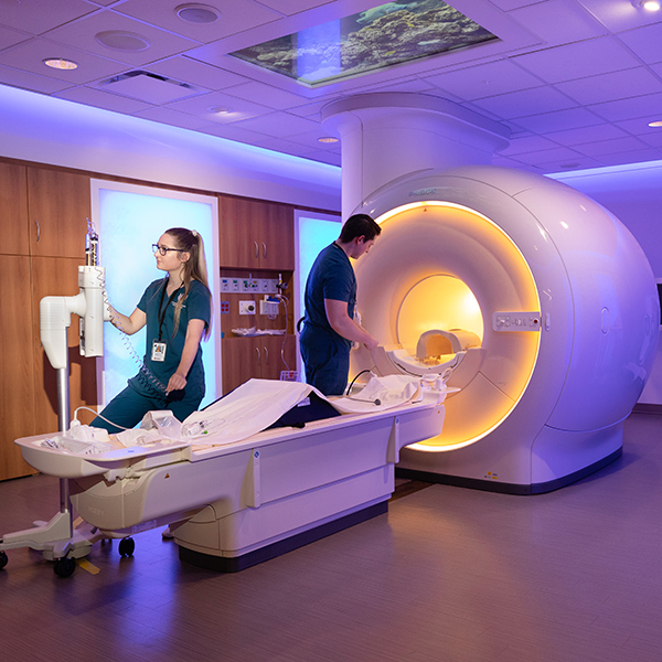
The MRI machine is a large, cylindrical (tube-shaped) machine that creates a strong magnetic field around the patient and sends pulses of radio waves from a scanner. Some MRI machines look like narrow tunnels, while others are more open.
The strong magnetic field created by the MRI scanner causes the atoms in your body to align in the same direction. Radio waves are then sent from the MRI machine and move these atoms out of the original position. As the radio waves are turned off, the atoms return to their original position and send back radio signals. These signals are received by a computer and converted into an image of the part of the body being examined. This image appears on a viewing monitor.
MRI may be used instead of computed tomography (CT) when organs or soft tissue are being studied. MRI is better at telling the difference between types of soft tissues and between normal and abnormal soft tissues.
Because ionizing radiation is not used, there is no risk of exposure to radiation during an MRI procedure.
Newer uses for MRI have contributed to the development of additional magnetic resonance technology. Magnetic resonance angiography (MRA) is a procedure used to evaluate blood flow through arteries. MRA can also be used to detect aneurysms in the brain and vascular malformations — abnormalities of blood vessels in the brain, spinal cord or other parts of the body.
Functional magnetic resonance imaging (fMRI) of the brain is used to determine the specific location in the brain where a certain function, such as speech or memory, occurs. The general areas of the brain in which such functions occur are known, but the exact location may vary from person to person. During fMRI of the brain, you will be asked to perform a specific task, such as reciting the Pledge of Allegiance. By pinpointing the exact location of the functional center in the brain, doctors can plan surgery or other treatments for a brain disorder.
How do I prepare for an MRI procedure?
EAT/DRINK: You may eat, drink and take medications as usual for most MRI exams. There are some specialty MRI exams that require certain restrictions. You will be provided detailed preparations instructions by Johns Hopkins Medical Imaging when you schedule your exam.
CLOTHING: You will be asked to remove all clothing, including underwear, and lock up all personal belongings. Please remove all piercings and leave all jewelry and valuables at home.
WHAT TO EXPECT: Imaging takes place inside of a large, tubelike structure that is open on both ends. You must lie perfectly still for quality images. Due to the MRI machine's loud noise, earplugs are required and will be provided.
ALLERGY: Some MRI exams require IV contrast. If you have had an allergic reaction to MRI contrast, contact your ordering physician to obtain the recommended prescription. You will likely take this by mouth 24, 12 and two hours prior to the examination.
ANTI-ANXIETY MEDICATION: If you require anti-anxiety medication due to claustrophobia, contact your ordering physician for a prescription. You must bring your prescription on the day of your appointment. Please note that you will need someone to drive you home.
STRONG MAGNETIC ENVIRONMENT: Due to the strong magnetic field, you must inform your doctor prior to the appointment if you have any metal in your body. Detailed information will be needed, such as the type and location, to determine your eligibility for MRI. If you have metal in your body that was not disclosed before your appointment, your study may be delayed, rescheduled or canceled upon your arrival until more information can be obtained.
Based on your medical condition, your health care provider may require other preparations.
When you call to make an appointment, it is extremely important that you inform your doctor if any of the following apply to you:
* You have a pacemaker or have had heart valves replaced.
* You have any type of implantable pump, such as an insulin pump.
* You have vessel coils, filters, stents, or clips.
* You are pregnant or think you might be pregnant.
* You have ever had a bullet wound.
* You have ever worked with metal (for example, as a metal grinder or welder).
* You have metallic fragments anywhere in the body.
* You are not able to lie down for 30 to 60 minutes.
How do I prepare for specialized MRI studies?
In some cases, you will be contacted prior to the examination to discuss the details of the procedure and how to prepare.
Specialized MRI exams include:
* breast MRI
* breast biopsy by MRI
* dynamic pelvis/defecography by MRI
* enterography by MRI
* functional MRI
* magnetic resonance angiography (MRA)
* prostate imaging by MRI
What happens during an MRI procedure?
MRI scans may be performed on an outpatient basis or as part of a stay in a hospital. Although specific protocols may differ among facilities, an MRI procedure generally follows this process:
* You will be asked to remove all clothing, jewelry, eyeglasses, hearing aids, hairpins, removable dental work or other objects that may interfere with the procedure.
* You will be given a gown to wear.
* If you are to have a procedure done with contrast, an IV line will be started in the hand or arm for injection of the contrast dye. If the contrast is to be taken by mouth, you will be given the contrast to swallow.
* You will lie on a scan table that slides into a large circular opening of the scanning machine. Pillows and straps may be used to prevent movement during the procedure.
* The technologist will be in another room where the scanner controls are located. However, you will be in constant sight of the technologist through a window. Speakers inside the scanner will enable the technologist to communicate with and hear you. You will have a communication ball so that you can let the technologist know if you have any problems during the procedure. The technologist will be watching you at all times and will be in constant communication.
* You will be given earplugs or a headset to help block out the noise from the scanner. Some headsets may play music.
* During the scanning process, a clicking noise will sound as the magnetic field is created and pulses of radio waves are sent from the scanner.
* It will be important for you to remain very still during the examination, as any movement could cause distortion and affect the quality of the scan.
* At intervals, you may be instructed to hold your breath or not breathe for a few seconds, depending on the body part being examined. You will then be told when you can breathe. You should not need to hold your breath for longer than a few seconds.
* If contrast dye is used, you may experience some effects when it is injected into the IV line. These effects include a flushing sensation or a feeling of coldness, a salty or metallic taste in the mouth, a brief headache, itching, or nausea and/or vomiting. These effects usually last for a few moments.
* You should notify the technologist if you have any breathing difficulties, sweating, numbness or heart palpitations.
* Once the scan is complete, the table will slide out of the scanner and you will be helped off the table.
* If an IV line was inserted for contrast administration, the line will be removed.
While the MRI procedure itself causes no pain, having to lie still for the length of the procedure might cause some discomfort or pain, particularly if you had a recent injury or invasive procedure, such as surgery. The technologist will use all possible comfort measures and complete the procedure as quickly as possible.
What happens after an MRI procedure?
You should move slowly when getting up from the scanner table to avoid any dizziness or lightheadedness from lying flat during the procedure.
If any sedatives were taken for the procedure, you may be required to rest until they have worn off. You will also need to avoid driving.
If contrast dye was used during the procedure and you experience any side effects or reactions to the contrast dye, such as itching, swelling, rash or difficulty breathing after your appointment call your doctor right away. If you feel it is a life threatening emergency, call 911.
If you notice any pain, redness and/or swelling at the IV site after you return home, you should notify your doctor, as this could indicate an infection or other type of reaction.
Otherwise, no special type of care is required after an MRI scan. You may resume your usual diet and activities unless your doctor advises you differently.
Your doctor may give you more or alternate instructions after the procedure depending on your particular situation.
Additional Information
An MRI (magnetic resonance imaging) scan is a test that creates clear images of the structures inside your body using a large magnet, radio waves and a computer. Healthcare providers use MRIs to evaluate, diagnose and monitor several different medical conditions.
What is an MRI?
An MRI (magnetic resonance imaging) scan is a painless test that produces very clear images of the organs and structures inside your body. MRI uses a large magnet, radio waves and a computer to produce these detailed images. It doesn’t use X-rays (radiation).
Because MRI doesn’t use X-rays or other radiation, it’s the imaging test of choice when people will need frequent imaging for diagnosis or treatment monitoring, especially of their brain.
What is an open MRI?
An open (or “open bore”) MRI refers to the type of machine that takes the images. Typically, an open MRI machine has two flat magnets positioned over and under you with a large space between them for you to lie. This allows for open space on two sides and alleviates much of the claustrophobia many people experience with closed-bore MRI machines.
However, open MRIs don’t take as clear images as closed-bore MRI machines. Closed-bore MRI machines have a ring of magnets that forms an open hole or tube in the middle where you’d lie to get the images. Closed-bore MRIs are narrow with tight head-to-ceiling space. This can cause anxiety and discomfort for some people, but these MRI machines take the best quality images.
If you’re nervous about your MRI scan or have a fear of closed spaces, talk to your healthcare provider. If needed, your provider will discuss options for sedatives (medicines to make you feel relaxed) or even anesthesia if necessary.
What is an MRI with contrast?
Some MRI exams use an injection of contrast material. The contrast agent contains gadolinium, which is a rare earth metal. When this substance is present in your body, it alters the magnetic properties of nearby water molecules, which enhances the quality of the images. This improves the sensitivity and specificity of the diagnostic images.
Contrast material enhances the visibility of the following:
* Tumors.
* Inflammation.
* Infection.
* Blood supply to certain organs.
* Blood vessels.
If your MRI requires a contrast material, a healthcare provider will insert an intravenous catheter (IV line) into a vein in your hand or arm. They’ll use this IV to inject the contrast material.
Contrast materials are safe drugs. Side effects ranging from mild to severe do occur, but severe reactions are very rare.
What’s the difference between an MRI scan and a CT scan?
Magnetic resonance imaging (MRI) uses magnets, radio waves and a computer to create images of the inside of your body, whereas computed tomography (CT) uses X-rays and computers.
Healthcare providers often prefer to use MRI scans instead of CT scans to look at the non-bony parts or soft tissues inside your body. MRI scans are also safer since they don’t use the damaging ionizing radiation of X-rays.
MRI scans also take much clearer pictures of your brain, spinal cord, nerves, muscles, ligaments and tendons than regular X-rays and CT scans.
However, not everyone can undergo an MRI. The magnetic field of MRI can displace metal implants or affect the function of devices such as pacemakers and insulin pumps. If this is the case, a CT scan is the next best option.
MRI scanning is usually more expensive than X-ray imaging or CT scanning.
What does an MRI show?
Magnetic resonance imaging (MRI) produces detailed images of the inside of your body. Healthcare providers can “look at” and evaluate several different structures inside your body using MRI, including:
* Your brain and surrounding nerve tissue.
* Organs in your chest and abdomen, including your heart, liver, biliary tract, kidneys, spleen, bowel, pancreas and adrenal glands.
* Breast tissue.
* Your spine and spinal cord.
* Pelvic organs, including your bladder and reproductive organs (uterus and ovaries in females and the prostate gland in males).
* Blood vessels.
* Lymph nodes.
When would I need an MRI?
Healthcare providers use magnetic resonance imaging (MRI) to help diagnose or monitor the treatment for many different conditions. There are also different types of MRIs based on which area of your body your provider wants to examine.
Brain and spinal cord MRIs can help evaluate and diagnose the following conditions:
* Brain aneurysms.
* Brain tumors and spinal tumors.
* Brain and spine injuries from trauma.
* Compression or inflammation of spinal cord and nerves (pinched nerve).
* Multiple sclerosis (MS).
* Spinal cord conditions.
* Spine anatomy and alignment.
* Stroke.
Providers use cardiac (heart) MRIs for several reasons, including:
* To evaluate the anatomy and function of your heart chambers, heart valves, size of and blood flow through major vessels and the surrounding structures.
* To diagnose cardiovascular conditions, such as tumors, infections and inflammatory conditions.
* To evaluate the effects of coronary artery disease, such as limited blood flow to your heart muscle and scarring within your heart muscle after a heart attack.
* To evaluate the anatomy and function of the heart and blood vessels in children and adults with congenital heart disease.
Body MRIs can evaluate structures and diagnose several conditions, including:
* Tumors in your chest, abdomen or pelvis.
* Liver diseases, such as cirrhosis, and issues with your bile ducts and pancreas.
* Inflammatory bowel disease, such as Crohn's disease and ulcerative colitis.
* Malformations of blood vessels and inflammation of the vessels (vasculitis).
* A developing fetus in your uterus.
MRIs of bones and joints can help evaluate:
* Bone infections (osteomyelitis).
* Bone tumors.
* Disk abnormalities in your spine.
* Joint issues caused by injuries.
Healthcare providers sometimes use breast MRIs with mammography to detect breast cancer, especially in people who have dense breast tissue or who might be at high risk of breast cancer.
Is an MRI safe?
An MRI scan is generally safe and poses almost no risk to the average person when appropriate safety guidelines are followed.
The strong magnetic field the MRI machines emit is not harmful to you, but it may cause implanted medical devices to malfunction or distort the images.
There’s a very slight risk of an allergic reaction if your MRI requires the use of contrast material. These reactions are usually mild and controllable by medication. If you have an allergic reaction, a healthcare provider will be available for immediate assistance.
Healthcare providers generally don’t perform gadolinium contrast-enhanced MRIs on pregnant women due to unknown risks to the developing fetus unless it’s absolutely necessary.
Who shouldn’t get an MRI?
In most cases, an MRI exam is safe for people with metal implants, except for a few types. Unless the device you have is certified as MRI safe, you might not be able to have an MRI. These devices may include:
* Metallic joint prostheses.
* Some cochlear implants.
* Some types of clips used for brain aneurysms.
* Some types of metal coils placed within blood vessels.
* Some older cardiac defibrillators and pacemakers.
* Vagal nerve stimulators.
If your healthcare provider recommends an MRI scan, they’ll ask detailed questions about your medical history and any medical devices or implants you may have in or on your body.
Test Details:
Who performs an MRI?
A radiologist or a radiology technologist will perform your MRI. A radiologist is a medical doctor who performs and interprets imaging tests to diagnose conditions. A radiology technologist is a healthcare provider who’s specially trained and certified to perform an MRI scan.
How does an MRI work?
Magnetic resonance imaging (MRI) works by passing an electric current through coiled wires to create a temporary magnetic field in your body. A transmitter/receiver in the machine then sends and receives radio waves. The computer then uses these signals to make digital images of the scanned area of your body.
What do I need to do to prepare for an MRI?
The magnetic resonance imaging (MRI) scanner uses strong magnets and radio wave signals that can cause heating or possible movement of some metal objects in your body. This could result in health and safety issues. It could also cause some implanted electronic medical devices to malfunction.
If you have metal-containing objects or implanted medical devices in your body, your healthcare provider needs to know about them before your MRI scan. Certain implanted objects may require additional scheduling arrangements and special instructions. Other items don’t require special instructions but may require an X-ray to check on the exact location of the object before your exam.
It appears to me that if one wants to make progress in mathematics, one should study the masters and not the pupils. - Niels Henrik Abel.
Nothing is better than reading and gaining more and more knowledge - Stephen William Hawking.
Offline
Pages: 1